SciELO - Scientific Electronic Library Online

 
vol.38 número3Análisis de ácidos grasos y esteroles del Líquen Everniopsis TrullaBiosorption of lead(ii) ions by dead bacterial Biomass isolated from mine water índice de autoresíndice de assuntospesquisa de artigos
Home Pagelista alfabética de periódicos  

Serviços Personalizados

Journal

Artigo

Indicadores

Links relacionados

Compartilhar


Revista Boliviana de Química

versão On-line ISSN 0250-5460

Rev. Bol. Quim vol.38 no.3 La Paz ago. 2021  Epub 31-Ago-2021

https://doi.org/10.34098/2078-3949.38.3.2 

SHORT REPORT

Synthesis and size estimation of silver nanoparticles, by reduction with aqueous extracts of calyces leaves and seeds of hibiscus sabdariffa linn: Promotion of green synthesis

Franklin Pacheco Coello1 

1School of Health Sciences, Department of Basic Sciences, Laboratory of Chemistry and Instrumental Analysis, Laboratory of Heavy Metals and Organic Solvents, Agro-Industrial Biotechnology Section of the "Dr. Francisco J. Triana Alonso" Biomedical Research Institute (BIOMED-UC) University of Carabobo, Postal Code 2103, phone +2446004000, Ruiz Pineda Street, La Morita II, Santa Rita Sector, Aragua State, Venezuela *Corresponding autor: fpacheco2@uc.edu.ve


ABSTRACT:

The synthesis of silver nanoparticles (AgNPs) has had a positive impact on biomedical sciences, thanks to its diverse biological potentialities, among which its antimicrobial activity and in vitro interaction with various chemotherapeutic drugs stand out. The study aimed at the synthesis of AgNPs, using aqueous extracts of calyces, leaves and seeds of an organic cultivation of Hibiscus sabdariffa and a solution of silver nitrate (AgNO3). The concentration of total phenolic compounds and flavonids was determined for each extract, by the methods of Folin-Ciocalteu and Marinova respectively, this with the purpose of guaranteeing the quality and content of compounds with reducing capacity. AgNPs synthesis conditions were optimized, referring to AgNO3 volume and concentration, extract volume (calyxes, leaves and seeds), pH, heating time and temperature, using the statistical program StatGraphics. The analyzes yielded a concentration of 17.42 ± 0.12 mg GAE / g (calyces), 9.03 ± 0.91 mg GAE / g (leaves) and 11.32 ± 0.36 mg GAE / g (seeds). The maximum absorption peaks were obtained at 424 nm for all the three extracts, with absorbances of 1.5240±0.32=30-32 nm (AgNPs-calyces), 0.5674±0.24=12-14 nm (AgNPs-leaves) and 0.764 ±0.18=18-20 nm (AgNPs-seeds). The findings of the study are related to the variation of phenolic compounds in the different parts of H. sabdariffa, which conditions the synthesis performance of AgNPs. The present study represents a valuable contribution in the "Green Synthesis", representing the first study in Venezuela in which AgNPs are synthesized from H. sabdariffa.

Keywords: Nanoparticles; Reduction; Hibiscus sabdariffa; Green Synthesis; Polyphenols.

RESUMEN:

La síntesis de nanopartículas de plata (AgNPs) ha tenido un impacto positivo en las ciencias biomédicas, gracias a sus diversas potencialidades bilógicas, entre las que destacan su actividad antimicrobiana e interacción in vitro con diversas drogas quimioterapéuticas. El estudio tuvo como objetivo la síntesis de AgNPs, empleando extractos acuosos de cálices, hojas y semillas de un cultivo orgánico de Hibiscus sabdariffa y una solución de nitrato de plata (AgNO3). A cada extracto se le determinó la concentración de compuestos fenólicos totales y flavonoides, por los métodos de Folin-Ciocalteu y Marinova respectivamente, esto con el propósito de garantizar la calidad y contenido de compuestos con capacidad reductora. Se optimizaron las condiciones de síntesis de AgNPs, referente a volumen y concentración de AgNO3, volumen de extracto (cálices, hojas y semillas), pH, tiempo y temperatura de calentamiento, empleando el programa estadístico StatGraphics. Los análisis arrojaron una concentración de 17.42±0.12 mg GAE/g (cálices), 9.03±0.91 mg GAE/g (hojas) y 11.32±0.36 mg GAE/g (semillas). Los picos máximos de absorción se obtuvieron a 424 nm para todos los tres extractos, con absorbancias de 1.5240±0.32 =30-32 nm (AgNPs- cálices), 0.5674±0.24=12-14 nm (AgNPs-hojas) y 0.7641±0.18=18-20 nm (AgNPs-semillas) . Los hallazgos del estudio están relacionados con la variación de compuestos fenólicos en las diferentes partes de H. sabdariffa, lo que condiciona el rendimiento de síntesis de AgNPs.el presente estudio representa un valioso aporte en la “Síntesis Verde”, representando el primer estudio en Venezuela en la que se sintetizan AgNPs a parir de H. sabdariffa.

Palabras clave: Nanopartículas; Reducción; Hibiscus sabdariffaa; Síntesis Verde; Polifenoles.

INTRODUCTION

Silver nanoparticles (AgNPs) have been one of the most attractive nanomaterials in biomedicine due to their unique physicochemical properties 1. Other biological activities of AgNPs have been also explored, including promoting bone healing and wound repair, enhancing the immunogenicity of vaccines, and anti-diabetic effects 2,3,4. Globally, research focus on the synthesis of AgNPs with controlled size and shape, and a variety of specific synthetic methods have been developed, including, chemical, physical and biological methods 5,6 (Figure 1).

Figure 1 Silver Nanoparticle System Mechanism. Source: Own elaboration 

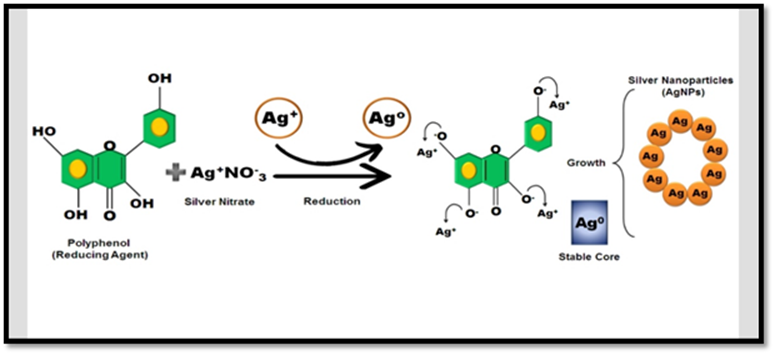
The synthesis of AgNPs due to biological processes aims to change the reagents used in chemical synthesis with another type of substances that can perform the same role. The "green synthesis" of silver nanoparticles, in which plant extracts are used, has gained great interest in recent years. This type of synthesis is efficient both in terms of reaction time, as well as stability of nanoparticles that exclude toxic chemical agents 7. In this sense Hibiscus sabdariffa, an easy-access and high-content plant in phenolic compounds, is an alternative as a bilingual material for AgNPs synthesis 8,9. Considering then the importance of AgNPs and their impact on biomedical sciences, the study aimed to synthesize and estimate the size of AgNPs, using aqueous extracts of calyxes, leaves and seeds of H. sabdariffa obtained from an organic culture, thus promoting the "Green Synthesis".

EXPERIMENTAL

Origin of plant material (PM)

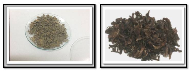
The calyces, leaves and seeds were obtained from an organic culture. The harvest was carried out on the researcher's own lands located in the Coropo sector, Aragua state, Venezuela (October 2019-March 2020).

Sample preparation for extraction

For extracts 2,5 grams of plant material were weighed. This was poured into a 400 mL Beaker, to which 200 mL of distilled water previously heated to the boiling point was added. The sample was slightly stirred for 4 min and filtered using Whatman No. 4 paper 10.

Determination of total phenolics

For the determination of total phenolics, 50 μL were mixed with 250 μL of the Folin-Ciocalteu 1 N reagent (Analytical grade, Merck). It was left to stand for 8 minutes and then 750 µL of 20% Na2CO3 and 950 µL of distilled water were added. Was incubated for 30 min at room temperature and the absorbance was read on a Genesis 20 UV/VIS spectrophotometer (Thermo Scientific, Waltham, Massachusetts, USA). A calibration curve for Gallic Acid (Sigma-Aldrich, Germany) was prepared with concentrations of 50, 100, 200, 300, 400, 500 and 1000 ppm. The results were expressed in mg of Gallic Acid Equivalents (GAE) /g of PM 11.

Figure 2 Leaves and calyces. 

Determination of flavonoids

A volume of 100 μL of sample was mixed with 30 μL of 5% w/v NaNO2, 30 μL of 10% w/v AlCl3, 200 μL of 1 M NaOH and adjusted with distilled water to a final volume of 1 mL. The reading was performed at 510 nm in a Genesis 20 UV / VIS spectrophotometer and was compared with a standard curve with standard (+)-catechin (Sigma Aldrich, USA). The results were expressed in mg of Catechin Equivalents (CE) / g of PM 12.

Synthesis of AgNPs-UV-VIS spectrophotometry

Nanoparticle formation was analyzed by UV-VIS Spectrophotometry (Thermo Scientific, Waltham, Massachusetts, USA). A spectral scan of each synthesis was carried out, obtained with the extracts of the various parts of the plant, in a range of wavelengths from 420 to 430 nm, estimating the size of the nanoparticles from (2 to 40 nm). For the analysis of the nanoparticles, 2 ml of each synthesis were taken and diluted with distilled water to a final volume of 4 mL. 1 mL of the previous dilution was placed in a cell with 1 cm of optical path and the spectral scan was carried out 13,14.

Optimisation of the synthesis of silver nanoparticles and Statistical analysis

The parameters to be optimised were: concentration of silver nitrate, volume of extract, pH, heating time and temperature. The StatGraphics programme was used with a screening design class. All determinations (phenolic compounds, flavonoids) and the spectral sweep corresponding to each synthesis were performed in quintuplicate and values were expressed as mean ± standard deviation. Statistical differences were determined by analysis of variance (ANOVA) using Statistic 9.0 for Windows.

RESULTS AND DISCUSSION

Total phenolics and flavonoids of the extracts

Prior to the synthesis and estimation of the size of the AgNPs, the concentration of total phenolics was determined in quintuplicate, obtaining a concentration of 17.42± 0.12 mg GAE / g PM (calyces), 9.03 ± 0.91 mg GAE/g PM (leaves) and 11.32±0.36 mg GAE/g PM (seeds), observing a statistical difference (p=0.035). For flavonoids it was of 12.17 ± 0.22 mg CE/g PM (calyces), 6.45 ± 0.67 mg CE/g PM (leaves) and 9.32 ± 0.28 mg CE/g PM (seeds) (p=0.045). The difference observed is similar to that reported in several studies carried out with Hibiscus sabdariffa, which indicate that the concentration of phenolic compounds and flavonoids it varies in the different parts of the plant, also conditioned by various factors (plant genetics, climate, soil conditions, cultivation) 15,16,17.

Optimal conditions for the synthesis of AgNPs

As indicated above, the material used corresponds to an organic culture of H. sabdariffa. In this sense, when using different parts of the pant, the appropriate conditions for the synthesis of nanoparticles were apotimized 18,19,20 (Table 1).

Table 1 Optimal conditions for the synthesis of AgNPs 

Optimization Calyces Leaves Seeds
*AgNO3 Solution (mL) 2.7 3.5 2.8
Time (min) 4.3 5.5 4.7
Temperature (°C) 91 82 84
Volume Extract (mL) +pH 4.7 7.3 6.8 6.6 8.0 7.1

*Silver nitrate concentration: 1.2 mM (Calices), 0.98 mM (leaves), 1.25 mM (Seeds). +pH adjusted with sodium hydroxide.

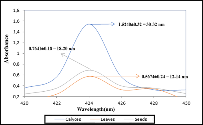
UV-VIS spectroscopy is a very powerful tool to monitor the synthesis of the AgNPs as the metallic nanoparticles possess a property known as surface plasmon resonance which is primarily because of the oscillation of the free electrons present on the surface of the metallic nanoparticles when they are excited by any external energy source1.The three solutions yielded maximum absorption peaks at 424 nm, observing that the synthesis of AgNPs, using the calyces, originated the highest absorbance value (Figure 3). All this shows that the characteristic peak for the formation of silver nanoparticles is around 422-426 nm, where the size of silver nanoparticles is estimated to be between 2 and 40 nm. 18,19.

On the other hand, the morphology of the synthesized nanoparticles plays a fundamental role in its biological activity21,22,23,24. Although the morphology of the AgNPs was not characterized, what was found in the study represents an important contribution to the green synthesis, leaving pending further studies, not only the characterization of the AgNPs, but also their biological potential.

Figure 3 Absorption spectrum of AgNPs. 

CONCLUSION

In conclusion, it must be said that the various parts of H. sabdariffa constitute an excellent alternative for the synthesis of silver nanoparticles, allowing to appreciate that its performance is conditioned to the presence of compounds with reducing capacity. Finally, this study provides interesting data for those researchers whose synthesis of nanoparticles is based on the "Green Synthesis".

REFERENCES

1. Li, X., Yi-Yi, W., Jie, H., Chun-Yuan, Ch., Zhen-Xing, W., Hui, X. 2020, Silver nanoparticles: Synthesis, medical applications and biosafety, Theranostics, 10(20), 8996-9031. DOI: 10.7150/thno.45413 [ Links ]

2. Mohandass, C., Vijayaraj, A.S., Rajasabapathy, R., Satheeshbabu, S., Rao, S.V., Shiva, C., De-Mello, I. 2013, Biosynthesis of Silver Nanoparticles from Marine Seaweed Sargassum cinereum and their Antibacterial Activity, Indian Journal Pharmaceutical Sciences, 75(5), 606-610, PMID: 24403664 [ Links ]

3. Asgary, V., Shoari, A., Baghbani-Arani, F., Sadat Shandiz, SA., Khosravy, MS., Janani, A., Bigdeli, R., Bashar, R., Cohan, RA. 2016, Green synthesis and evaluation of silver nanoparticles as adjuvant in rabies veterinary vaccine, International Journal of Nanomedicine, 11, 3597-3605. DOI: https://doi.org/10.2147/IJN.S109098Links ]

4. Saratale, G.D., Saratale, R.G., Benelli, G., Kumar, G., Pugazhendhi, A., Kim, D-S., Shin, H-S. 2017, Anti-diabetic potential of silver nanoparticles synthesized with Argyreia nervosa leaf extract high synergistic antibacterial activity with standard antibiotics against foodborne bacteria, Journal of Cluster Science, 28(3), 1709-1727. DOI: https://doi.org/10.1007/s10876-017-1179-zLinks ]

5. Shanmuganathan, R., Karuppusamy, I., Saravanan, M., Muthukumar, H., Ponnuchamy, K., Ramkumar, VS., Pugazhendhi, A. 2019, Synthesis of Silver Nanoparticles and their Biomedical Applications - A Comprehensive Review, Current Pharmaceutical Design, 25(24), 2650-2660. DOI: 10.2174/1381612825666190708185506 [ Links ]

6. Roy, S., Anantharaman, P. 2018, Biosynthesis of Silver Nanoparticle by Amphiroa anceps (Lamarck) Decaisne and Its Biomedical and Ecological Implications, Journal of Nanomedicine & Nanotechnology, 9(2), 487- 492. DOI: 10.4172/2157-7439.1000492 [ Links ]

7. Okafor, F., Janen, A., Kukhtareva, T., Edwards, V., Curley, M. 2013, Green synthesis of silver nanoparticles, their characterization, application and antibacterial activity, International journal of environmental research and public health, 10(10), 5221-5238. DOI: https://doi.org/10.3390/ijerph10105221Links ]

8. Ghazala, R., Rajni, C. 2018, A review on phytochemistry and therapeutic uses of Hibiscus sabdariffa L., Biomedicine & Pharmacotherapy, 102, 575-586. DOI: https://doi.org/10.1016/j.biopha.2018.03.023Links ]

9. Pacheco Coello, F., Orosco-Vargas, C., Peraza-Marrero, M., Pinto-Catari, I., Ramirez-Azuaje, D. 2020, Effect of an extract of Hibiscus sabdariffa L., on oxidative stress induced in Saccharomyces cerevisiae, Ciencia, Ambiente y Clima, 3(1), 41-46. DOI: https://doi.org/10.22206/cac.2020.v3i1.pp41-46Links ]

10. Reyes-Luengas, A., Salinas-Moreno, Y., Ovando- Cruz, M., Arteaga-Garibay, R. 2015, Análisis de ácidos fenólicos y actividad antioxidante de extractos acuosos de variedades de jamaica (Hibiscus Sabdariffa L.) con cálices de colores diversos, Agrociencia, 49,277-290 [ Links ]

11. Ayala-Zavala, JF., Silva-Espinoza, A.B., Cruz-Valenzuela, R.M., Villegas-Ochoa, M.A. 2013, Antioxidant and antifungal potential of metanol extracts of Phenillus sp, Revista Iberoaméricana de Micolología, 29(3), 132-138 [ Links ]

12. Marinova, D., Ribarova, F., Atanassova, M. 2005, Total phenolics and total flavonoids in bulgarian fruits and vegetables, Journal of the University of Chemical Technology and Metallurgy, 40(3), 255-260 [ Links ]

13. Rodríguez-León, E., Iñiguez-Palomares, R., Navarro, R. E., Herrera-Urbina, R., Tánori, J., Iñiguez-Palomares, C., Maldonado, A. 2013, Synthesis of silver nanoparticles using reducing agents obtained from natural sources (Rumex hymenosepalus extracts), Nanoscale Research Letters, 8(1), 318. DOI: https://doi.org/10.1186/1556-276X-8-318Links ]

14. Kannan, R.R.R., Stirk, W.A., Van Staden, J. 2013, Synthesis of silver nanoparticles using the seaweed Codium capitatum P. C. Silva (Chlorophyceae), South African Journal of Botany, 86, 1-4. DOI: https://doi.org/10.1016/j.sajb.2013.01.003Links ]

15. Juhari, N.H., Bredie, W.L.P., Toldam-Andersen, T.B., Petersen, M.A. 2018, Characterization of Roselle calyx from different geographical origins, Food Research International, 12, 378-389. DOI: 10.1016/j.foodres.2018.06.049 [ Links ]

16. Ramirez-Azuaje, D., Pinto-Catari, I., Peraza-Marrero, M., Orosco-Vargas, C., Pacheco-Coello, F. 2018, Hibiscus sabdariff L. Una comparación de compuestos fenólicos totales y flavonoides en cálices y hoja, VITAE, 76,1-5 [ Links ]

17. Idowu-Adebayo, F., Toohey, M.J., Fogliano, V., Linnemann, A.R. 2021, Enriching street-vended zobo (Hibiscus sabdariffa) drink with turmeric (Curcuma longa) to increase its health-supporting properties, Food and Function, 12(2), 761-770. DOI: https://doi.org/10.1039/D0FO02888FLinks ]

18. Wiley, B.J., Im, S.H., Li, Z.Y., McLellan, J., Xia, Y. 2006, Maneuvering the surface plasmon resonance of silver nanostructures through shape-controlled synthesis, The Journal of Physical Chemistry B, 110(32), 15666-15675. DOI: https://doi.org/10.1021/jp0608628Links ]

19. Dubey, S.P., Lahtinen, M., Sillanpää, M. 2010, Green synthesis and characterizations of silver and gold nanoparticles using leaf extract of rosa rugosa, Colloids and Surfaces A: Physicochemical and Engineering Aspects, 364(1-3 ), 34-41. DOI: https://doi.org/10.1016/j.colsurfa.2010.04.023Links ]

20. Nayak, D., Ashe, S., Rauta, PR., Nayak, B. 2015, Biosynthesis, characterization and antimicrobial activity of silver nanoparticles using Hibiscus rosa-sinensis petals extracts, Institution of Engineering and Technology Nanobiotechnol, 9(5), 288-93. DOI: https://doi.org/10.1049/iet-nbt.2014.0047Links ]

21. Koli, S.H., Mohite, B.V., Suryawanshi, R.K., Borase, H.P., Patil, S.V. 2018, Extracellular red Monascus pigment-mediated rapid one-step synthesis of silver nanoparticles and its application in biomedical and environment, Bioprocess and Biosystems Engineering, 41(5), 715-727. DOI: https://doi.org/10.1007/s00449-018-1905-4Links ]

22. Mao, B.H., Chen, Z.Y., Wang, Y.J., Yan, S.J. 2018, Silver nanoparticles have lethal and sublethal adverse effects on development and longevity by inducing ROS-mediated stress responses, Scientific Reports, 8(2445), 1-16. DOI: https://doi.org/10.1038/s41598-018-20728-zLinks ]

23. Naaz, S., Poddar, S., Bayen, S.P., Mondal, M.K., Roy, D., Mondal, S.K., Chowdhury P., Saha, S.K. 2018, Tenfold enhancement of fluorescence quantum yield of water soluble silver nanoclusters for nano-molar level glucose sensing and precise determination of blood glucose level, Sensors and Actuators. B, Chemical, 255(Part 1), 332-340. DOI: https://doi.org/10.1016/j.snb.2017.07.143Links ]

24. Akther, T., Mathipi, V., Kumar, N.S, Davoodbasha, M., Srinivasan, H. 2019, Fungal-mediated synthesis of pharmaceutically active silver nanoparticles and anticancer property against A549 cells through apoptosis, Environmental science and Pollution Research International, 26, 13649-13657. DOI: https://doi.org/10.1007/s11356-019-04718-wLinks ]

Received: March 24, 2021; Accepted: August 21, 2021; Published: August 30, 2021

Creative Commons License This is an open-access article distributed under the terms of the Creative Commons Attribution License