




 English (pdf)
 Article in xml format
 Article references

 Automatic translation
 Send this article by e-mail
 Cited by SciELO
 Similars in
SciELO
 uBio

 Permalink

INTRODUCTION
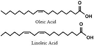
Chemically, edible oils are composed of 98% triglycerides (glycerol esterified with three fatty acids) with different substitution patterns, chain lengths and degrees of unsaturation, and some minor components (2%) such as sterols, phospholipids, carotenes and fat-soluble vitamins (vitamins A, D, E and K), sugars and more complex components that can be released by numerous hydrolytic reactions. Edible oils are good sources of nutrition for the human diet, which are necessary for the proper development of human tissues. The main chemical molecules in vegetable oils are oleic acid and linoleic acid (Fig. 1).
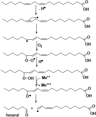
The phenomenon of fatty acid rancidity is technically known as oxidative degradation or lipid peroxidation, and is one of the parameters that most affects the quality of fatty products1. Lipids tend to auto-oxidation and oxidation processes such as the frying process (160 - 190 °C) in the presence of oxygen and humidity, these conditions favor the formation of lipid hydroperoxide radicals which are compounds that are formed during the oxidation of lipids, this process is called lipid peroxidation. Hydroperoxide radicals react with fatty acids and generate peroxides which, when they reach their maximum value, are converted into aldehydes, ketones and short-chain fatty acids, forming a typical rancid aroma and flavor, called secondary products of lipid oxidation2, the general mechanism is shown in figure 2.3 The consequences of these chemical substances are very significant for health, which when absorbed by food and subsequently by the body negatively affect health2.
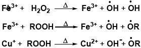
The main factors in the rancidity process are: The presence of metallic ions, such as iron and copper, which act as catalysts in various stages of the peroxidation process, many of the cooking processes are generally carried out on this type of metals (Fig. 3).
Exposure to temperatures above 70 °C in the presence of oxygen significantly accelerates the peroxidation process; this occurs mainly during frying, as seen in Figure 4.
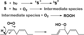
There are two categories of sensitizing photoreceptors. The first, exemplified by riboflavin (vitamin B2), becomes activated in a triplet excited state (³S*) when exposed to light and interacts directly with unsaturated fatty acids and their esters. This interaction forms an intermediate compound that subsequently reacts with molecular oxygen (O₂), resulting in the formation of hydroperoxides and the return of the sensitizer to its ground state (S)1.
Photosensitizers of the second type1, like chlorophyll in its unexcited form (S), absorb light and first enter an excited singlet state (¹S), then shift to a triplet excited state (³S). While in this triplet state and still exposed to light (hν), they react with molecular oxygen (O₂), producing an intermediate. This intermediate, along with another sensitizer molecule in the singlet excited state (¹S*), leads to the formation of singlet oxygen (ΔG ¹O₂)-a highly reactive form of oxygen that directly attacks fatty acids or their esters (RH), resulting in the production of hydroperoxides (ROOH), see Fig. 4.
Prolonged exposure to radiant energy sources, such as solar radiation, causes the initiation or propagation of lipid peroxidation, a situation observed in fried foods that come in transparent packaging (potato chips in polyethylene packaging) or photosensitizing substances such as chlorophyll (Figure 5). The presence of antioxidants, such as carotenes and vitamin E, inhibits, or slow, the process of lipid peroxidation.

The main organoleptic molecules that are responsible for rancidity are; 3-nonenal, 2,4-decadienal, acetaldehyde, acrolein, propanal, butanal, pentanal, heptanal, nonanal, 2,3-hexenal, 2-heptenal, 2-octenal, 2,4-nonadienal, methyl 8-oxooctanoate, methyl 9-oxononanoate, malondialdehyde, methyl 10-oxodecanoate, methyl 10-oxo-8-decenoate, 4,5-epoxy-2-decenal, 4-hydroxy-trans-nonenal, 4-hydroxy-trans-hexanal, acrotonaldehyde, pentane, hexane, 1-pentanol, 1-octen-3-ol, 2-pentylfuran, methyl heptanoate and methyl octanoate.4
The molecules produced by lipid peroxidation that are most risky to health are malondialdehyde, acrolein, 4-hydroxy-trans-nonenal, 4-hydroxy-trans-hexanal and compounds similar to croton aldehyde. These substances are also involved in the pathogenesis of many diseases such as inflammatory processes, aging, cancer, arteriosclerosis and Alzheimer's disease5,6,7.
Lipid hydroperoxides contribute to cellular cytotoxicity, while aldehydes and oxysterols have proinflammatory, cytotoxic and mutagenic effects8. The end products of oxidation have been considered to cause inflammation, fibrosis and atypical cell proliferation7.
Oils oxidized by heating, their consumption could increase the risk of cardiovascular disease, endothelial dysfunction, lipid peroxidation, oxidative stress, genotoxicity, carcinogenicity and reduced glucose uptake. Furthermore, excessive generation of free radicals is known to cause liver damage, resulting in diseases such as hepatitis, cirrhosis, and liver tumors9. The formation of trans fatty acids in reused oils is harmful because it decreases good cholesterol and increases bad cholesterol, in addition to increasing triglyceride concentrations and the risk of heart attack and cancer10. Indeed, the toxicological and pathogenic properties that could result from the ingestion of lipid oxidation products, such as aldehydes contained in cooking oils heated according to standard frying practices, influence the potential development of cardiovascular diseases, carcinogenic properties, contribution to neurodegenerative disorders, hypertensive effects, development of diabetes, and respiratory and pulmonary complications, especially in the case of acrolein11,12.
Acrylamide, lipid oxidation, is one of the main chemical reactions occurring during food processing and can have a strong impact on the final quality of food. A significant role for carbonyl compounds derived from lipid oxidation in the formation of acrylamide has recently been proposed13,14. The mechanism of acrylamide formation is generally explained by the Maillard reaction, where the dominant interactions occur between reducing sugars or carbonyl compounds such as aldehydes or ketones and amino acids, especially asparagine (present in different types of meat). Acrylamide is widely known as a human neurotoxin, and low doses of acrylamide (0.5 mg/kg body weight/day) have been reported to be genotoxic and carcinogenic to animals15.
For the analysis of rancidity determination, peroxide analysis the following methods are used: Sensory analysis, Determination of the peroxide value, p-Anisidine analysis, Determination of the TBARS (Thiobarbituric Acid Reactive Substances) value, Kreis method, Gas chromatography coupled with mass spectrometry (GC-MS).
FTIR spectroscopy can be considered a "green chemical analysis method" because it reduces the use of toxic chemical reagents, is easy to use, rapid, sensitive, requires no sample preparation, and has been widely used in food sample analysis. It is one of the widely used vibrational spectroscopies for the characterization of all aspects of edible oils. IR spectroscopy can be defined as the interaction between electromagnetic radiation in the IR region in the form of scattering, reflection, absorption, or transmission with analyzed substances based on wavenumbers. The frequencies or wavelengths at which samples absorb IR radiation and their corresponding intensities (either transmittance or absorbance) are recorded in an IR spectrum. IR spectroscopy can be considered an ideal technique for the analysis of edible oils because this technique is rapid, easy in sample presentation, non-destructive, and non-invasive, which means that samples analyzed with IR spectroscopy can be analyzed using different instruments.
Currently, Fourier transform infrared spectroscopy (FTIR) has emerged as a potential tool for assessing the effects of degradation in vegetable oils. Spectral trends show the occurrence of oxidation, with reduction of unsaturation, in addition to the fractionation of carbon chains and the appearance of functional groups such as aldehydes and alcohols16. Fourier transform infrared spectroscopy (FTIR), combined with chemometrics, has been widely used to determine acid, peroxide, and saponification values. FTIR has been used to determine the acid value in edible oils by -O-H stretching, the peroxide value in edible oils by -COO stretching, and to monitor the peroxide value in oxidized emulsions17. NIR (Near-Infrared) spectroscopy can scan the absorbance or reflectance of molecules in the region between 800 and 2500 nm. This method has become popular for assessing the quality of food-related bioorganic materials because it does not require toxic organic solvents and involves rapid measurement. Some researchers have applied NIR spectroscopy for the differentiation and determination of edible fats and oils18,19.
One of the most significant bands in oxidized oils is the one associated with the C-O sigma bond, typically observed between 1100 and 1300 cm⁻¹, indicating the presence of ethers, peroxides, and acids. As the oil oxidizes, this band tends to intensify, while the bands associated with double bonds (C=C, =C-H) decrease. RO-O groups (in hydroperoxides) can appear between 850 and 888 cm⁻¹, although these vibrations are weaker, corresponding to aliphatic RO-O peroxides20,21,22.
The peroxide value can be correlated with specific spectroscopic signals, thus offering a rapid and direct monitoring alternative in quality control laboratories. In this context, the present study aims to determine the presence of lipid peroxides in edible oils collected from fast-food outlets using infrared spectroscopy. Specifically, it seeks to identify the presence of C-O bonds associated with oxidation products by comparing samples of used oils with a fresh control. Likewise, IR spectroscopy has been applied to predict acrylamide content in fried food samples, especially in French fries, and has been confirmed as a promising methodology. The work carried out with FT-IR showed, through its analysis, that the range 1490 cm-1-1699 cm-1 corresponds to the N-H vibrations.
RESULTS AND DISCUSSION
Table 1 shows the organoleptic evaluation of the analyzed samples, recording characteristics such as color, viscosity, and odor. These aspects are related to the oxidation levels observed in the FT-IR spectra.

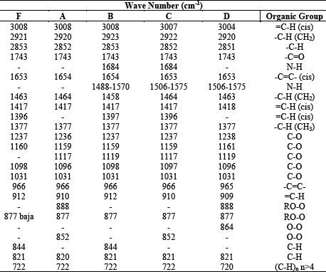
Table 2 presents IR-FT absorption band values for the reference sample (F) and samples A, B, C, D in wavenumbers (cm⁻¹).
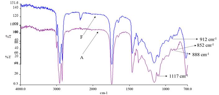
In figure 6, the main signals that differentiate oil A and F are observed. The IR spectrum of oil F shows a signal at 912 cm-1 that is attributed to vinyl C-H vibrations; this signal is not observed at 912 cm-1 in oil A. In oil A, C-O vibrations can be seen at 1117 cm-1, a signal that is not observed in oil F. Finally, in oil A, two signals are observed at 852 cm-1 and 888 cm-1 that correspond to aliphatic peroxide vibrations RO-O20 21.
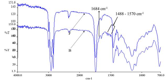
Figure 7 shows a difference between oil F and B in the region between 1488 cm-1 and 1570 cm-1 corresponding to N-H vibrations. Signals of oxidized carbons C-O and C=O are observed between 1031 cm -1 and 1237 cm-1, mainly a signal is observed at 1117 cm-1 due to O-C-O oxidation cleavages, an aspect that is not observed in oil F.
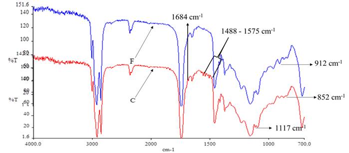
In Figure 8, the difference between oils F and C is in the region between 1488 cm-1 and 1575 cm-1 these vibrations are attributed to N-H of sample C. Another important signal of oil C is at 852 cm-1 which is attributable to aliphatic peroxides RO-O, on the other hand, the absence of the signal at 912 cm-1 in C is shown attributable to the aliphatic C-H and vinyl vibrations, the vibrations of the oxygenated carbons C-O and C=O are observed at 1117 cm-1.
In Figure 9, spectrum D shows vibrations between 1506 cm-1 and 1575 cm-1 that are attributed to N-H vibrations. Other important signals that D presents are at 852 cm-1 and 864 cm-1 attributable to aliphatic peroxides RO-O, also at 1031 cm-1 a signal is observed that corresponds to C-O vibrations. Signals at 1119 cm-1 of oil D are attributed to vibrations of oxygenated carbons C-O and C=O. All the signals mentioned above are not present in sample F. On the other hand, the absence in D of the vinyl =C-H vibrations at 912 cm-1.
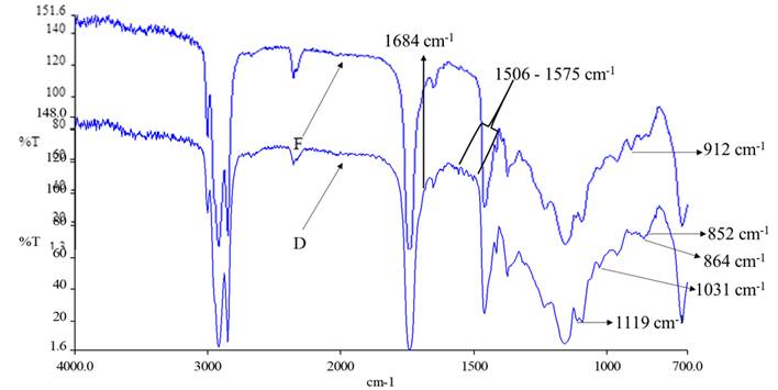
Figure 10 shows the summary of the spectra of samples A, B, C, D and F. In the region between 1488 cm-1 and 1575 cm-1 N-H vibration signals are shown in B, C and D, which are absent in F and A. In the region between 1117 cm-1 and 1119 cm-1, signals are observed in A, B, C and D that are attributed to C-O vibrations, signals that are not observed in oil F.
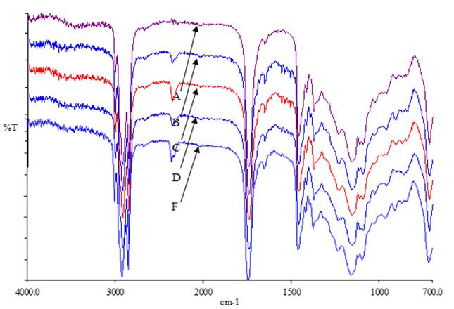
According to Silverstein20 Rao21 and Conley22, signals between the region of 875 cm-1 - 855 cm-1 correspond to RO-O stretching frequencies attributed to aliphatic peroxides. In oil A two signals are observed at 852 cm-1 and 888 cm-1, oil C one signal at 852 cm-1, oil D one signal at 864 cm-1, due to the above observations, the presence of the RO-O stretching vibration corresponding to aliphatic peroxides in oil samples A, C and D can be mentioned.
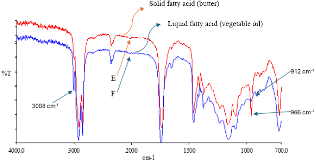
Figure 11 shows the IR spectra of sample E (lard) and sample F (unused oil). In both cases, a common signal is observed at 3008 cm⁻¹, corresponding to the =C-H stretching of unsaturated fatty acids; however, this band is less intense in butter due to its lower degree of unsaturation. A notable difference occurs in the 966 cm⁻¹ region, where sample E presents an intense band, absent in oil F. This signal is attributed to out-of-plane vibrations of trans vinyl bonds, or to vibration modes characteristic of saturated crystalline structures, typical of solid fats such as butter. In contrast, in the spectrum of oil F, a signal is distinguished at 912 cm⁻¹, corresponding to the vibration of the =C-H bonds in vinyl carbons, indicative of the presence of unsaturated fatty acids. This signal does not appear in butter, reinforcing its predominantly saturated composition. These spectral differences allow a clear distinction between saturated and unsaturated fatty acids. Based on the above, we can conclude that the locations where the samples were obtained do not use butter (E).
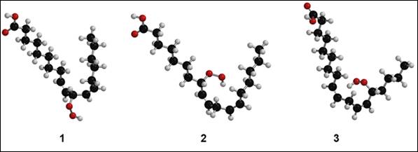
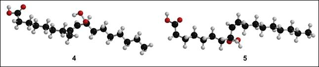
The computational calculation was performed in Equilibrium Geometry, with Density Functional Theory (DFT), EDF2/6-31G* (Spartan 18), which allowed the calculation of three isomers of linoleic acid and two of oleic acid, which includes the O-O group in alpha positions with respect to the double bonds as shown in Figures 12 and 13.
The calculations confirmed the vibrations at 855 cm-1 asymmetric bending vibration of 4 (C-O-O), 854 cm-1 stretching vibration of 1 (O-O), 875 cm-1 bending vibration of 1 (C-O-O), 927 cm-1 antisymmetric stretching vibration of 1 (C-O-O), 871 cm-1 bending vibration of 5 (C-O-O), 877 cm-1 bending vibration of 3 (C-O-O), 925 cm-1 stretching vibration of 3 (O-O), 902 cm-1 symmetric stretching of 4 (O-O), 891 cm-1 asymmetric vibration of 5 (C-O-O), 921 cm-1 stretching vibration of 5 (O-O), 887 cm-1 stretching vibration of 1 (O-O), 827 cm-1 bending vibration of 2 (C-O-O), 898 cm-1 symmetric stretching vibration of 2 (C-O-O), 928 cm-1 antisymmetric stretching vibration of 2, 928 cm-1 antisymmetric stretching vibration of 5 (C-O-O). This analysis allows us to confirm that the different isomers with respect to the peroxide, showing us vibrations between 855 cm-1 and 928 cm-1 for O-O and C-O-O bonds.
EXPERIMENTAL
Sample Collection
Four samples of used frying oil were collected from different food stalls located in the city of El Alto, La Paz, Bolivia (4,000 m above sea level). Sample A was obtained from roast beef; sample B was obtained from preparing chicken cracklings at a street stall; sample C was collected from a spit-roasted chicken restaurant; sample D was a darker, more viscous oil recovered from a store where repeated use of the same oil was observed; sample E was solid fat (lard); and sample F was unused edible vegetable oil, used as a reference for comparative analyses. Each sample was transferred to glass containers stored at room temperature and protected from light until analysis. The samples were characterized by both organoleptic inspection (color, viscosity, and appearance) and FT-IR spectroscopic analysis.
CONCLUSIONS
The signals shown in A, B, C, and D are similar to those in F, indicating that the collection sites use oil in cooking. This is confirmed by the F spectrum, which corresponds to butter. All samples exhibit rancid aromas. From the analysis of the samples, samples B, C, and D show N-H vibration signals between the 1488 cm-1 and 1575 cm-1 regions, attributed to acrylamide or derivatives that exhibit N-H vibrations. In samples A, C, and D, signals are observed between 852 cm-1 and 888 cm-1, corresponding to O-O and RO-O vibrations, corresponding to aliphatic peroxides. The computational calculation Density Functional Theory (DFT), EDF2/6-31G* (Spartan 18), allowed us to accurately understand the vibration signals of the O-O, RO-O, and N-H bonds with the experimental data of IR-FT and confirmed the presence of oils in the form of peroxides, acrylamide, and nitrogen derivatives, which shows a high degradation and/or oxidation of the evaluated samples. This confirms that the collected samples are harmful to health.

