<?xml version="1.0" encoding="ISO-8859-1"?><article xmlns:mml="http://www.w3.org/1998/Math/MathML" xmlns:xlink="http://www.w3.org/1999/xlink" xmlns:xsi="http://www.w3.org/2001/XMLSchema-instance">
<front>
<journal-meta>
<journal-id>2310-0265</journal-id>
<journal-title><![CDATA[Revista CON-CIENCIA]]></journal-title>
<abbrev-journal-title><![CDATA[Rev.Cs.Farm. y Bioq]]></abbrev-journal-title>
<issn>2310-0265</issn>
<publisher>
<publisher-name><![CDATA[Facultad de Ciencias Farmacéuticas y BioquímicasUniversidad Mayor de San Andrés]]></publisher-name>
</publisher>
</journal-meta>
<article-meta>
<article-id>S2310-02652020000200003</article-id>
<title-group>
<article-title xml:lang="en"><![CDATA[Structural dynamics between the open and closed conformations of the K ATP channel in pancreatic cells]]></article-title>
<article-title xml:lang="es"><![CDATA[Dinámica estructural entre la conformación abierta y cerrada del canal K ATP en células pancreáticas]]></article-title>
</title-group>
<contrib-group>
<contrib contrib-type="author">
<name>
<surname><![CDATA[Ballon Paucara]]></surname>
<given-names><![CDATA[Wendy Guadalupe]]></given-names>
</name>
<xref ref-type="aff" rid="A01"/>
</contrib>
<contrib contrib-type="author">
<name>
<surname><![CDATA[Grados-Torrez]]></surname>
<given-names><![CDATA[Ricardo Enrique]]></given-names>
</name>
<xref ref-type="aff" rid="A02"/>
</contrib>
</contrib-group>
<aff id="A01">
<institution><![CDATA[,Universidad Mayor de San Andrés Instituto de Investigaciones Fármaco Bioquímicas  Luis Enrique Terrazas Siles ]]></institution>
<addr-line><![CDATA[ ]]></addr-line>
</aff>
<aff id="A02">
<institution><![CDATA[,Universidad Mayor de San Andrés Instituto de Investigaciones Fármaco Bioquímicas  Luis Enrique Terrazas Siles ]]></institution>
<addr-line><![CDATA[ ]]></addr-line>
</aff>
<pub-date pub-type="pub">
<day>00</day>
<month>11</month>
<year>2020</year>
</pub-date>
<pub-date pub-type="epub">
<day>00</day>
<month>11</month>
<year>2020</year>
</pub-date>
<volume>8</volume>
<numero>2</numero>
<fpage>21</fpage>
<lpage>36</lpage>
<copyright-statement/>
<copyright-year/>
<self-uri xlink:href="http://www.scielo.org.bo/scielo.php?script=sci_arttext&amp;pid=S2310-02652020000200003&amp;lng=en&amp;nrm=iso"></self-uri><self-uri xlink:href="http://www.scielo.org.bo/scielo.php?script=sci_abstract&amp;pid=S2310-02652020000200003&amp;lng=en&amp;nrm=iso"></self-uri><self-uri xlink:href="http://www.scielo.org.bo/scielo.php?script=sci_pdf&amp;pid=S2310-02652020000200003&amp;lng=en&amp;nrm=iso"></self-uri><abstract abstract-type="short" xml:lang="en"><p><![CDATA[Abstract Introduction: The ATP-sensitive Potassium channel (K ATP channel) regulates insulin production by pancreatic &#946; cells. Glibenclamide (GBM) (antidiabetic drug) and ATP act as inhibitors of this channel, while ADP activates it. The K ATP channel is an octamer consisting of 4 central Kir6.2 subunits that form the pore and 4 external regulation subunits SUR1. Objective: To determine the structural dynamics between the open and closed conformations of the K ATP channel in pancreatic cells. Method: Comparative structural analysis of different crystallographic structures of the K ATP channel of human pancreatic cells using Chimera v1.11.2. Results: The Kir6.2 subunit has a PIP2 binding domain (activator), an Interfacial Helix (IFH) and an N-terminal domain (KNtp). On the other hand, the SUR1 subunit that contains the GBM binding site, has 2 Nucleotide Binding Domains (NBD1/2), an M5-Lh1 loop and a Lasso Motif formed by the interface between the Trans-membrane Domain 0 and Loop 0 (TMD0-L0). The results of the dynamic structural analysis using bioinformatics tools indicate that these regions participate actively in the conformational changes that lead to the closure (inhibition) or opening (activation) of this channel. Conclusion: The study of the dynamics of activation and inhibition of the K ATP channels is essential for the evaluation, discovery and/or design of new natural compounds, which like GBM, can promote insulin secretion to aid or improve the treatment of diabetic patients.]]></p></abstract>
<abstract abstract-type="short" xml:lang="es"><p><![CDATA[Resumen Introducción: El canal de Potasio sensible a ATP (canal K ATP) regula la producción de Insulina por células &#946; pancreáticas. La Glibenclamida (GBM) (fármaco antidiabético) y el ATP actúan como inhibidores de este canal, mientras que el ADP lo activa. El canal K ATP es un octámero constituido por 4 subunidades centrales Kir6.2 que forman el poro y 4 subunidades externas de regulación SUR1. Objetivo: Determinar la dinámica estructural entre las conformaciones abierta y cerrada del canal K ATP en células pancreáticas. Método: Análisis estructural comparativo de diferentes estructuras cristalográficas del canal K ATP de células pancreáticas humanas empleando el software Chimera v1.11.2. Resultados: La subunidad Kir6.2 presenta un dominio de unión a PIP2 (activador), una Hélice Interfacial (IFH) y un dominio N-terminal (KNtp). Por otro lado, la subunidad SUR1 que contiene el sitio de unión a la GBM, tiene 2 Dominios de Unión a Nucleótidos (NBD1/2), un bucle M5-Lh1 y un Motivo de Lazo formado por la interface entre el Dominio Trans-membrana 0 y el Bucle 0 (TMD0-L0). Los resultados del análisis dinámico estructural mediante herramientas bioinformáticas, indican que estas regiones participan activamente en los cambios conformacionales que dan lugar al cierre (inhibición) o apertura (activación) de este canal. Conclusión: El estudio de la dinámica de activación e inhibición de los canales K ATP es imprescindible para la evaluación, descubrimiento y/o diseño de nuevos compuestos naturales, que como la GBM, puedan promover la secreción de Insulina para coadyuvar o mejorar el tratamiento de pacientes diabéticos]]></p></abstract>
<kwd-group>
<kwd lng="en"><![CDATA[K ATP Channel]]></kwd>
<kwd lng="en"><![CDATA[Structural Dynamics]]></kwd>
<kwd lng="en"><![CDATA[Kir6.2]]></kwd>
<kwd lng="en"><![CDATA[SUR1]]></kwd>
<kwd lng="en"><![CDATA[Glibenclamide]]></kwd>
<kwd lng="en"><![CDATA[ATP and ADP]]></kwd>
<kwd lng="es"><![CDATA[Canal K ATP]]></kwd>
<kwd lng="es"><![CDATA[Dinámica Estructural]]></kwd>
<kwd lng="es"><![CDATA[Kir6.2]]></kwd>
<kwd lng="es"><![CDATA[SUR1]]></kwd>
<kwd lng="es"><![CDATA[Glibenclamida]]></kwd>
<kwd lng="es"><![CDATA[ATP]]></kwd>
<kwd lng="es"><![CDATA[ADP]]></kwd>
</kwd-group>
</article-meta>
</front><body><![CDATA[ <p align=right><font size="2" face="Verdana, Arial, Helvetica, sans-serif"><strong>ART&Iacute;CULOS ORIGINALES</strong></font></p>     <p align=right>&nbsp;</p>     <p align=center><font size="4" face="Verdana, Arial, Helvetica, sans-serif"><strong>Structural dynamics between the open and   closed conformations of     <br>   the K<sub>ATP</sub> channel in pancreatic cells</strong></font></p>     <p align=center>&nbsp;</p>     <p align=center><font size="3" face="Verdana, Arial, Helvetica, sans-serif"><strong>Dinámica estructural entre la conformación abierta y cerrada del     <br> canal K<sub>ATP </sub>en células pancreáticas</strong></font></p>      <p align="center">&nbsp;</p>     <p align="center">&nbsp;</p>     <p align="center"><font size="2" face="Verdana, Arial, Helvetica, sans-serif"><strong>Ballon Paucara Wendy Guadalupe<sup>1</sup><a href="" target="_self" onClick="javascript: w = window.open('https://orcid.org/0000-0001-8088-8000','','width=640,height=500,resizable=yes,scrollbars=1,menubar=yes,');"><img src="/img/revistas/rcfb/v8n2/id_orcid.png" width="16" height="16" border="0"></a></strong>, <strong>&nbsp;Grados-Torrez Ricardo Enrique<sup>1*</sup><a href="" target="_self" onClick="javascript: w = window.open('https://orcid.org/0000-0001-7287-2586','','width=640,height=500,resizable=yes,scrollbars=1,menubar=yes,');"><img src="/img/revistas/rcfb/v8n2/id_orcid.png" width="16" height="16" border="0"></a></strong></font>    ]]></body>
<body><![CDATA[<br> <font size="2" face="Verdana, Arial, Helvetica, sans-serif"><sup>1 </sup>Area de Farmacología, Instituto de Investigaciones Fármaco Bioquímicas ”Luis Enrique Terrazas Siles”. Universidad Mayor     <br> de San Andrés, Av. Saavedra 2224. La Paz, Bolivia.</font>    <br> <font size="2" face="Verdana, Arial, Helvetica, sans-serif">*Author for correspondence</font>: <font size="2" face="Verdana, Arial, Helvetica, sans-serif">Wendy Guadalupe Ballon Paucara</font> <font size="2" face="Verdana, Arial, Helvetica, sans-serif">Licenciada en Bioquímica.     <br> Unidad de Bioquímica Clínica, Área de Farmacología. Instituto de Investigaciones Fármaco Bioquímicas “Luis Enrique     <br> Terrazas Siles”  Facultad de Ciencias Farmacéuticas y Bioquímicas. Universidad Mayor de San Andrés.</font> <font size="2" face="Verdana, Arial, Helvetica, sans-serif">    <br> E-mail: <a href="mailto:wen.ballon.wen@gmail.com">wen.ballon.wen@gmail.com</a></font>    <br> <font size="2" face="Verdana, Arial, Helvetica, sans-serif">Principal author:</font> <font size="2" face="Verdana, Arial, Helvetica, sans-serif">Ricardo Enrique Grados Torrez (Author for correspondence).</font>    <br> <font size="2" face="Verdana, Arial, Helvetica, sans-serif">PhD en Biotecnología, MSc. en Biotecnología Molecular, Lic. en Bioquímica. Área de Farmacología, Instituto de     <br> Investigaciones   Fármaco Bioquímicas (I.I.F.B.) ”Luis Enrique Terrazas Siles”. Universidad Mayor de San Andrés,     <br> Av. Saavedra 2224.   La Paz, Bolivia.</font>    ]]></body>
<body><![CDATA[<br> <font size="2" face="Verdana, Arial, Helvetica, sans-serif">E-mail: <a href="mailto:ric.grados@gmail.com">ric.grados@gmail.com</a></font> <font size="2" face="Verdana, Arial, Helvetica, sans-serif">Cel. 65192791</font>    <br> <font size="2" face="Verdana, Arial, Helvetica, sans-serif"><b>Fecha de recepci&oacute;n: </b>22 Septiembre 2020<b>       Fecha de aceptaci&oacute;n: </b>12 Octubre de 2020</font></p>      <p align="justify">&nbsp;</p>     <p align="justify">&nbsp;</p> <hr>     <p align="justify"><font size="2" face="Verdana, Arial, Helvetica, sans-serif"><b>Abstract</b></font></p>     <p align=justify><font size="2" face="Verdana, Arial, Helvetica, sans-serif"><b>Introduction:</b> The ATP-sensitive   Potassium channel (K<sub>ATP</sub> channel) regulates insulin production by   pancreatic &#946; cells. Glibenclamide (GBM) (antidiabetic drug) and ATP act as   inhibitors of this channel, while ADP activates it. The K<sub>ATP</sub> channel   is an octamer consisting of 4 central Kir6.2 subunits that form the pore and 4   external regulation subunits SUR1.</font></p>     <p align="justify"><font size="2" face="Verdana, Arial, Helvetica, sans-serif"><b>Objective: </b>To determine the structural dynamics between the open and closed conformations of the K<sub>ATP</sub> channel in pancreatic cells.</font></p>      <p align="justify"><font size="2" face="Verdana, Arial, Helvetica, sans-serif"><b>Method: </b>Comparative structural analysis of different crystallographic structures of the K<sub>ATP</sub> channel of human pancreatic cells using Chimera v1.11.2.</font></p>      <p align="justify"><font size="2" face="Verdana, Arial, Helvetica, sans-serif"><b>Results:</b> The Kir6.2 subunit has a PIP<sub>2</sub> binding domain (activator), an Interfacial Helix (IFH) and an N-terminal domain (KNtp). On the other hand, the SUR1 subunit that contains the GBM binding site, has 2 Nucleotide Binding Domains (NBD1/2), an M5-Lh1 loop and a Lasso Motif formed by the interface between the Trans-membrane Domain 0 and Loop 0 (TMD0-L0). The results of the dynamic structural analysis using bioinformatics tools indicate that these regions participate actively in the conformational changes that lead to the closure (inhibition) or opening (activation) of this channel.</font></p>      <p align="justify"><font size="2" face="Verdana, Arial, Helvetica, sans-serif"><b>Conclusion: </b>The study of the dynamics of activation and inhibition of the K<sub>ATP</sub> channels is essential for the evaluation, discovery and/or design of new natural compounds, which like GBM, can promote insulin secretion to aid or improve the treatment of diabetic patients. </font></p>      ]]></body>
<body><![CDATA[<p align="justify"><font size="2" face="Verdana, Arial, Helvetica, sans-serif"><b>Keywords: </b>K<sub>ATP</sub> Channel, Structural Dynamics, Kir6.2, SUR1, Glibenclamide, ATP and ADP.</font></p> <hr>     <p align="justify"><font size="2" face="Verdana, Arial, Helvetica, sans-serif"><b>Resumen</b></font></p>     <p align="justify"><font size="2" face="Verdana, Arial, Helvetica, sans-serif"><b>Introducción: </b>El   canal de Potasio sensible a ATP (canal K<sub>ATP</sub>) regula la producción de   Insulina por células &#946; pancreáticas. La Glibenclamida (GBM) (fármaco   antidiabético) y el ATP actúan como inhibidores de este canal, mientras que el   ADP lo activa. El canal K<sub>ATP</sub> es un octámero constituido por 4   subunidades centrales Kir6.2 que forman el poro y 4 subunidades externas de   regulación SUR1. </font></p>     <p align="justify"><font size="2" face="Verdana, Arial, Helvetica, sans-serif"><b>Objetivo: </b>Determinar la dinámica estructural entre las conformaciones abierta y cerrada del canal K<sub>ATP</sub> en células pancreáticas.</font></p>      <p align="justify"><font size="2" face="Verdana, Arial, Helvetica, sans-serif"><b>Método: </b>Análisis estructural comparativo de diferentes estructuras cristalográficas del canal K<sub>ATP</sub> de células pancreáticas humanas empleando el software Chimera v1.11.2.</font></p>      <p align="justify"><font size="2" face="Verdana, Arial, Helvetica, sans-serif"><b>Resultados:</b> La subunidad Kir6.2 presenta un dominio de unión a PIP<sub>2 </sub>(activador), una Hélice Interfacial (IFH) y un dominio N-terminal (KNtp). Por otro lado, la subunidad SUR1 que contiene el sitio de unión a la GBM, tiene 2 Dominios de Unión a Nucleótidos (NBD1/2), un bucle M5-Lh1 y un Motivo de Lazo formado por la interface entre el Dominio Trans-membrana 0 y el Bucle 0 (TMD0-L0). Los resultados del análisis dinámico estructural mediante herramientas bioinformáticas, indican que estas regiones participan activamente en los cambios conformacionales que dan lugar al cierre (inhibición) o apertura (activación) de este canal. </font></p>      <p align="justify"><font size="2" face="Verdana, Arial, Helvetica, sans-serif"><b>Conclusión: </b>El estudio de la dinámica de activación e inhibición de los canales K<sub>ATP</sub> es imprescindible para la evaluación, descubrimiento y/o diseño de nuevos compuestos naturales, que como la GBM, puedan promover la secreción de Insulina para coadyuvar o mejorar el tratamiento de pacientes diabéticos.</font></p>      <p align="justify"><font size="2" face="Verdana, Arial, Helvetica, sans-serif"><b>Palabras clave: </b>Canal K<sub>ATP</sub>, Dinámica Estructural, Kir6.2, SUR1, Glibenclamida, ATP y ADP</font></p> <hr>     <p align="justify">&nbsp;</p>     <p align="justify">&nbsp;</p>     ]]></body>
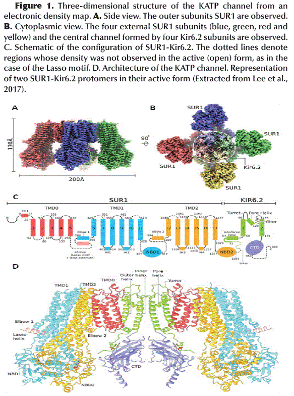
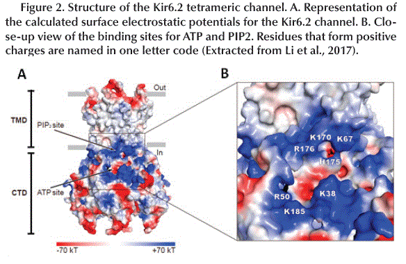
<body><![CDATA[<p align="justify"><font size="3" face="Verdana, Arial, Helvetica, sans-serif"><b>INTRODUCTION</b></font></p>     <p align="justify"><font size="2" face="Verdana, Arial, Helvetica, sans-serif">Insulin controls the concentration of Glucose in the blood. When this concentration is high, a group of pancreatic &#946; cells release this hormone. In these cells, the amount of sugar in the blood modifies the ATP/ADP ratio and a membrane protein called ATP-sensitive Potassium channel (K<sub>ATP</sub> channel) acts as a switch that activates or deactivates the production of Insulin. ATP and ADP control this switch, since both have opposite effects on this channel (ATP act as inhibitor and ADP as activator) (Aittoniemi <i>et al.,</i> 2009), therefore, their action is coupled with the release of Insulin (Aguilar-Bryan <i>et al.,</i> 2001; Ashcroft, 2005). However, it is not yet clear how K<sub>ATP</sub> channels detect changes in the ATP/ADP ratio in these cells, and because ATP levels are usually high and constant, ATP is continuously deactivating these channels while it is still unclear how ADP activates those (Lee <i>et al., </i>2017). On the other hand, Glibenclamide (GBM), sulfonylurea used in the conventional treatment of Type 2 Diabetes Mellitus (T2DM), also inhibits K<sub>ATP</sub> channels promoting Insulin secretion. Understanding the conformational changes that occur in the K<sub>ATP </sub>channel is important for the discovery of new natural compounds that promote Insulin secretion in patients with T2DM.</font></p>     <p align="justify"><font size="2" face="Verdana, Arial, Helvetica, sans-serif"><b>Structure of K<sub>ATP</sub> channel</b></font></p>     <p align="justify"><font size="2" face="Verdana, Arial, Helvetica, sans-serif">Functional   K<sub>ATP</sub> channels are membrane proteins that have a hetero-octameric   structure composed of four Internal Rectification Subunits (Kir6.2) that close   in depolarization maintaining the membrane potential and four Sulfonylurea   Receptor Subunits (SUR1) members of the ABCC family (ATP Binding Cassette,   subfamily C) (Lee <i>et al., </i>2017) (<a href="#f1">Figure 1A</a> and <a href="#f1">B</a>), with a total of 80   trans-membrane helices (3 helices for each Kir6.2 subunit and 17 helices for   each SUR1 subunit) (<a href="#f1">Figure 1C</a>) and with a total molecular weight of about 880   kDa (Li <i>et al., </i>2017; Martin <i>et al., </i>2017b). The K<sub>ATP</sub>   channel pore is made up of Kir6.2 subunits that act as ATP sensors and their   activity is regulated by PIP<sub>2</sub>. The SUR1 subunits mediate the   activating effect of ADP-Mg<sup>2+</sup> and determine the pharmacological   profile of the channel (Wu <i>et al., </i>2018). It should be noted that the K<sub>ATP</sub>   channels have different combinations of subunits and different pharmacological   profiles in different tissues, but the specific K<sub>ATP</sub> channels of   pancreatic &#946; cells are mainly composed of Kir6.2 and SUR1 subunits and are   highly sensitive to anti diabetic drugs such as GBM (Ashcroft, 2005; Martin <i>et     al., </i>2019).</font></p>     <p align="center"><a name="f1"></a><img src="/img/revistas/rcfb/v8n2/a03_figura01.gif" width="583" height="784"></p>     <p align="justify"><font size="2" face="Verdana, Arial, Helvetica, sans-serif"><b>Kir6.2</b></font></p>     <p align="justify"><font size="2" face="Verdana, Arial, Helvetica, sans-serif">The   tetrameric structure of Kir6.2 subunits forms the channel itself and has   two-layer architecture, a Trans-Membrane Domain (TMD) and a Globular Cytosolic   Domain (CTD) (<a href="#f2">Figure 2A</a>). The TMD has three trans-membrane helices: the Pore   Helix (PH), an Inner Helix (IH or M2) that connects to the CTD and one the   Outer Helix (OH or M1) that connects to the cytoplasm with a small Interfacial   Helix (IFH) which has a final extension in the form of a &#946;A-IFH loop (<a href="#f1">Figure 1C</a> and <a href="#f1">D</a>), this loop   is connected to the N-terminal region (KNtp) which is highly dynamic (Li <i>et     al., </i>2017).</font></p>     <p align="center"><font size="2" face="Verdana, Arial, Helvetica, sans-serif"><a name="f2"></a><img src="/img/revistas/rcfb/v8n2/a03_figura02.gif" width="584" height="380"></font></p>     <p align="justify"><font size="2" face="Verdana, Arial, Helvetica, sans-serif">The surface electrostatic potential shows two positively charged regions. The first consists of K67, K170, H175 and R176 which is known as the PIP<sub>2 </sub>binding site which is an activator of K<sub>ATP</sub> channels (open the channel). The second region is formed by R50, K38 and K185 located near the cavity that accommodates the Adenosine group of ATP (ATP binding site) (Hansen <i>et al.,</i> 2011) (<a href="#f2">Figure 2B</a>).</font></p>     <p align="justify"><font size="2" face="Verdana, Arial, Helvetica, sans-serif"><b>SUR1</b></font></p>     ]]></body>
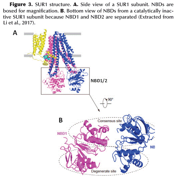
<body><![CDATA[<p align="justify"><font size="2" face="Verdana, Arial, Helvetica, sans-serif">The   Kir6.2 tetrameric channel is surrounded by four SUR1 subunits. Each divided   into a TMD0 (with 5 trans-membrane helices: M1-5) with adjacent cytosolic loop   L0 (Loop 0) and an ABC transporter-like domain TMD1-TMD2 (with 12   trans-membrane helices: TMD1 M6-11 and TMD2 M12-17) (Aittoniemi <i>et al.,</i>   2009) (<a href="#f1">Figure 1C</a>). SUR1 TMD1-TMD2 has an inward-facing conformation since the   central cavity is completely accessible to the cytosolic face. The interface   formed between TMD0-L0 and TMD1-TMD2 is called the Lasso Motif (<a href="#f1">Figure 1C</a> and   <a href="#f1">D</a>). Inwardly, TMD1 and TMD2 possess a Nucleotide Binding Domain NBD1 and NBD2,   respectively (<a href="#f3">Figure 3A</a>), which form a catalytic site for ATP hydrolysis when   both NBDs acquire a dimeric conformation (one close to the other) (Zhang and   Chen, 2016). ABC-like domains have a characteristic motif (LSGGQ) and a   Glutamate (E855). However, in SUR1 the NBDs are functionally asymmetric, since   NBD1 presents an Aspartate instead of Glutamate (E855D) and NBD2 does not   retain the characteristic ABC motif, therefore, one site is consensus and the   other is degenerated (Vedovato <i>et al.,</i> 2015) (<a href="#f3">Figure 3B</a>).</font></p>     <p align="center"><a name="f3"></a><img src="/img/revistas/rcfb/v8n2/a03_figura03.gif" width="586" height="604"></p>     <p align="justify">&nbsp;</p>     <p align="justify"><font size="3" face="Verdana, Arial, Helvetica, sans-serif"><b>METHOD</b></font></p>     <p align="justify"><font size="2" face="Verdana, Arial, Helvetica, sans-serif">In this study, a comparative structural analysis of different crystallographic structures of the K<sub>ATP</sub> channel of human pancreatic cells was performed, obtained from the Protein Data Bank database (PDB: <a href="http://www.rcsb.org/" target="_blank">http://www.rcsb.org/</a>).</font></p>      <p align="justify"><font size="2" face="Verdana, Arial, Helvetica, sans-serif"><b>6baa: </b>Crystallographic structure of K<sub>ATP</sub> channel from pancreatic &#946; cells bound to ATP and GBM, this structure was used for the analysis of the GBM binding site.</font><font size="2" face="Verdana, Arial, Helvetica, sans-serif"><b>&nbsp;</b></font></p>      <p align="justify"><font size="2" face="Verdana, Arial, Helvetica, sans-serif"><b>5ykf: </b>Crystallographic structure of K<sub>ATP</sub> channel from pancreatic &#946; cells bound to ATP&#947;S and GBM, this structure was used for the analysis of conformational changes in SUR1 and the NBD1 and NBD2 domains when the channel is in its closed conformation.</font></p>      <p align="justify"><font size="2" face="Verdana, Arial, Helvetica, sans-serif"><b>5ywc: </b>Crystallographic structure of the K<sub>ATP</sub> channel from pancreatic &#946; cells bound to ADP-Mg<sup>2+</sup>, this structure was used for the analysis of conformational changes in SUR1 and the NBD1 and NBD2 domains when the channel is in its open conformation.</font><font size="2" face="Verdana, Arial, Helvetica, sans-serif"><b>&nbsp;</b></font></p>      <p align="justify"><font size="2" face="Verdana, Arial, Helvetica, sans-serif"><b>6pza: </b>Crystallographic structure of the SUR1 subunit from pancreatic &#946; cells bound to ATP and GBM, this structure was used for the analysis of the Lasso Motif (TMD0-L0) and the N-terminal domain of Kir6.2 (KNtp).</font></p>      <p align="justify"><font size="2" face="Verdana, Arial, Helvetica, sans-serif">All the structures were analyzed, evaluated and modeled using Chimera v1.11.2 software. Superposition and structural alignments were made using the Match Maker tool with Iteration no greater than 2 Å.</font></p>     ]]></body>
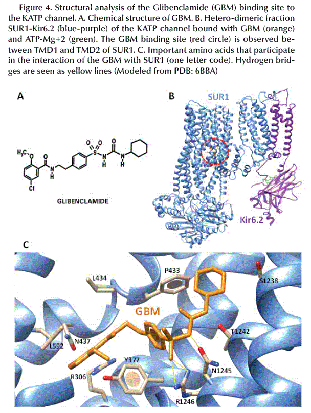
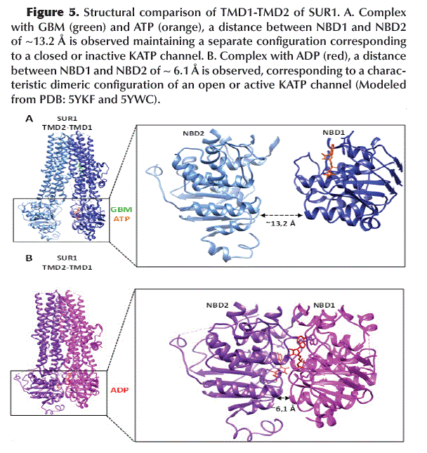
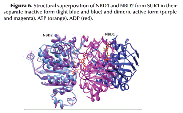
<body><![CDATA[<p align="justify">&nbsp;</p>     <p align="justify"><font size="3" face="Verdana, Arial, Helvetica, sans-serif"><b>RESULTS</b></font><font size="2" face="Verdana, Arial, Helvetica, sans-serif"><b>&nbsp;</b></font></p>     <p align="justify"><font size="2" face="Verdana, Arial, Helvetica, sans-serif"><b>Binding site of GBM</b></font></p>     <p align="justify"><font size="2" face="Verdana, Arial, Helvetica, sans-serif">The   chemical structure of GBM (<a href="#f4">Figure 4A</a>) allows it to interact to the SUR1 subunit   between the TMD1 and TMD2 domains in the K<sub>ATP</sub> channel (<a href="#f4">Figure 4B</a>).   The amino acids involved in this interaction are mainly aromatic and   hydrophobic such as Y377, P434, L434 and L592 and form hydrogen bonds between   R1246/N1245 and -SO2/-NH- (<a href="#f4">Figure 4C</a>).</font></p>     <p align="center"><a name="f4"></a><img src="/img/revistas/rcfb/v8n2/a03_figura04.gif" width="608" height="808"></p>     <p align="justify"><font size="2" face="Verdana, Arial, Helvetica, sans-serif"><b>NBD1-NBD2 dimerization</b></font><font size="2" face="Verdana, Arial, Helvetica, sans-serif"><b>&nbsp;</b></font></p>     <p align="justify"><font size="2" face="Verdana, Arial, Helvetica, sans-serif">Binding of GBM (with SUR1) and ATP (with NBD1) keeps the NBD1 and NBD2 domains (located at TMD1 and TMD2, respectively) in a separate configuration (~13.2 Å apart) corresponding to a closed or inactive K<sub>ATP</sub> channel (<a href="#f5">Figure 5A</a>). In contrast, binding of ADP to NBD1 and NBD2 promotes its dimerization, with both domains closest to each other (~6.1 Å apart) which is characteristic of an open or active K<sub>ATP </sub>channel (<a href="#f5">Figure 5B</a>).</font></p>      <p align="center"><a name="f5"></a><img src="/img/revistas/rcfb/v8n2/a03_figura05.gif" width="608" height="645"></p>     <p align="justify"><font size="2" face="Verdana, Arial, Helvetica, sans-serif">The superposition of the   structures of NBD1 and NBD2 indicates an evident conformational change induced   by the binding of GBM/ATP or ADP (<a href="#f6">Figure 6</a>).</font></p>     <p align="center"><a name="f6"></a><img src="/img/revistas/rcfb/v8n2/a03_figura06.gif" width="595" height="369"></p>      ]]></body>
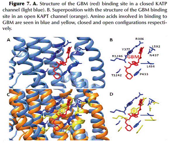
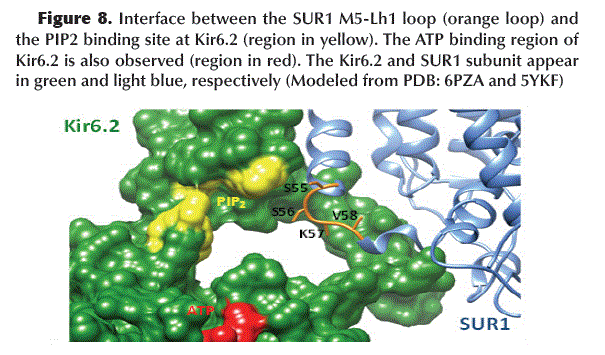
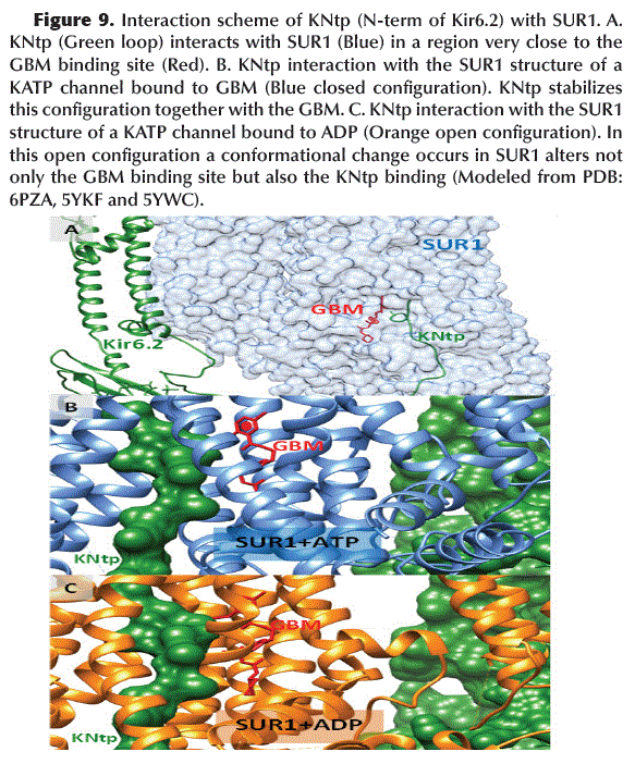
<body><![CDATA[<p align="justify"><font size="2" face="Verdana, Arial, Helvetica, sans-serif"><b>Occlusion of GBM binding site</b></font></p>      <p align="justify"><font size="2" face="Verdana, Arial, Helvetica, sans-serif">After dimerization of NBD1 and NBD2 by the binding of ADP, a conformational change in SUR1 results in the narrowing of the GBM binding site, primarily by rotation of the Y377 and P433 residues (<a href="#f7">Figure 7</a>), which maintains the K<sub>ATP</sub> channel in its active or open configuration.</font></p>     <p align="center"><a name="f7"></a><img src="/img/revistas/rcfb/v8n2/a03_figura07.gif" width="602" height="491"></p>     <p align="justify"><font size="2" face="Verdana, Arial, Helvetica, sans-serif"><b>Binding site of PIP<sub>2</sub> is blocked</b></font></p>     <p align="justify"><font size="2" face="Verdana, Arial, Helvetica, sans-serif">The   binding of GBM to SUR1 induces a conformational change in the M5-Lh1 loop (S55,   S56, K57 and V58) of TMD1-SUR1 acquiring a configuration that blocks the PIP<sub>2</sub>   binding site located in Kir6.2 (<a href="#f8">Figure 8</a>).</font></p>     <p align="center"><a name="f8"></a><img src="/img/revistas/rcfb/v8n2/a03_figura08.gif" width="605" height="355"></p>      <p align="justify"><font size="2" face="Verdana, Arial, Helvetica, sans-serif"><b>Role of KNtp in the activation of K<sub>ATP</sub> channel</b></font><font size="2" face="Verdana, Arial, Helvetica, sans-serif"><b>&nbsp;</b></font></p>      <p align="justify"><font size="2" face="Verdana, Arial, Helvetica, sans-serif">The N-terminal region of Kir6.2 (KNtp) interacts inside SUR1 very close to the GBM binding site (<a href="#f9">Figure 9A</a>). Apparently, this interaction stabilizes the structure of the K<sub>ATP</sub> channel in a closed (or inactive) configuration (<a href="#f9">Figure 9B</a>), whereas, the dimerization of NBD1 and NBD2 (located in TMD1 and 2 of SUR1, respectively) after ADP binding, it causes a conformational change in SUR1 that is incompatible with the stabilizing interaction of KNtp and the binding of GBM in SUR1 (<a href="#f9">Figure 9C</a>) causing the opening of the K<sub>ATP </sub>channel.</font></p>      <p align="center"><a name="f9"></a><img src="/img/revistas/rcfb/v8n2/a03_figura09.gif" width="583" height="702"></p>     <p align="justify"><font size="2" face="Verdana, Arial, Helvetica, sans-serif"><b>L0 and IFH communicate SUR1 with Kir6.2</b></font></p>     ]]></body>
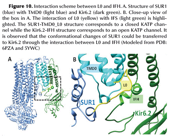
<body><![CDATA[<p align="justify"><font size="2" face="Verdana, Arial, Helvetica, sans-serif">The   L0 loop present in the TMD0 of SUR1 in its inactive state (closed K<sub>ATP</sub>   channel) acquires a configuration that allows it to interact with the Kir6.2   IFH (Interface Loop) (<a href="#f10">Figure 10</a>). This interaction probably promotes the Kir6.2   subunits (which shape the K<sub>ATP</sub> channel pore) to acquire a closed   configuration.</font></p>     <p align="center"><font size="2" face="Verdana, Arial, Helvetica, sans-serif"><a name="f10"></a><img src="/img/revistas/rcfb/v8n2/a03_figura10.gif" width="584" height="474"></font></p>     <p align="justify">&nbsp;</p>     <p align="justify"><font size="3" face="Verdana, Arial, Helvetica, sans-serif"><b>DISCUSSION</b></font></p>     <p align="justify"><font size="2" face="Verdana, Arial, Helvetica, sans-serif">GBM   is located in the interface between TMD0-L0 and TMD1-TMD2 acting as a wedge to   stabilize the SUR1 structure in an inactive or closed conformation (with NBD1   and 2 separated) and also stabilizing the TMD0-L0 fragment in a conformation   that could inhibit the opening of Kir6.2 (Li <i>et al., </i>2017). A   comparative study points out that in the presence of GBM and ATP-Mg<sup>2+</sup>   both the internal and external helices of the Kir6.2 TMD move inward giving   rise to a channel with a narrow and closed helix input (channel inactive or closed).</font></p>     <p align="justify"><font size="2" face="Verdana, Arial, Helvetica, sans-serif">All   Kir6.2 residues that interact with SUR1 are located primarily before the   helical structures that make up the pore of the K<sub>ATP</sub> channel, the   Kir6.2 fragments composed of the outer helices such as IFH and the N-terminal   region (KNtp) they are sufficient to interact with SUR1. The Lasso Motif   (present in the L0 interface) could act as a region that communicates the   signal sent by the SUR1 regulatory subunits to the Kir6.2 subunits that are the   ones that make up the channel <i>per se</i>, since in closed conformation, this   motif interacts with IFH (Kir6.2 Interfacial Loop) possibly promoting a   conformational change that results in the closure of the K<sub>ATP</sub>   channel. However, the Lasso Motif is only visible in the inactive (closed) but   not in the active (open) configuration due to the low resolution and low   electron density obtained in the crystallographic structures and the great   mobility that this region has (Martin <i>et al., </i>2019). Other studies   indicate that when the K<sub>ATP</sub> channel is in its active (open) form   (called the &quot;quatrefoil&quot; state), the Lasso Motif acquires a   disordered configuration representing a decoupled state between SUR1 and   Kir6.2, in which the conformational change of SUR1 cannot be transferred to Kir6.2 or vice versa (Lee <i>et al., </i>2017).</font></p>     <p align="justify"><font size="2" face="Verdana, Arial, Helvetica, sans-serif">Other   studies based on the microscopic technique of reconstruction of molecules on an   atomic scale (Lee <i>et al., </i>2017), allowed obtaining 3D reconstruction   maps for the K<sub>ATP</sub> channel of human pancreatic cells. The maps show   that these channels have binding sites for ATP and ADP. The ADP binding site   acts as a sensor that can detect small changes in cellular levels of ADP. In   addition, a dynamic Loop-like structure was revealed that connects the ATP and   ADP junction areas, this Motif plays an important role allowing the increase in   ADP to cancel the action that ATP has on the K<sub>ATP</sub> channel (since ADP   and ATP have opposite effects). The presence of an ADP sensor and the Lasso   Motif could explain how K<sub>ATP</sub> channels monitor changes in the ATP/ADP   ratio and can therefore control the release of Insulin based on blood Glucose   levels. Genetic defects in the functioning of the K<sub>ATP</sub> channels of   the pancreas can cause many diseases (Nichols, 2006; Quan <i>et al.,</i> 2011).   Understanding the structure and function of these channels is essential for the   discovery, analysis, or design of new drugs that serve to complement or improve   the treatment of diseases such as T2DM.</font></p>     <p align="justify">&nbsp;</p>     <p align="justify"><font size="3" face="Verdana, Arial, Helvetica, sans-serif"><b>CONCLUSION</b></font></p>     <p align="justify"><font size="2" face="Verdana, Arial, Helvetica, sans-serif">The   results obtained from the comparison of different crystallographic structures   of the K<sub>ATP</sub> channel from human pancreatic cells, support the   activation model proposed by Wu <i>et al.,</i> 2018, in which both GMB and KNtp   (N-terminal Kir6.2) bind cooperatively within the SUR1 subunit and inhibit activation   (opening) of the K<sub>ATP</sub> channel, while ATP may be linked to NBD1   (degenerate site) but with separate NBD1/2 domains. In turn, the M5-Lh1 loop of   SUR1 blocks the PIP<sub>2</sub> binding site (which is a K<sub>ATP</sub>   channel activator) located at Kir6.2. When the ADP concentration increases, it   first binds to NBD2 (consensus site) inducing a conformational change and   increasing the affinity of NBD1 for ADP or ATP, promoting the closure and   dimerization of both NBDs. Finally, a global conformational change of SUR1 occurs   (due to NBD1/2 dimerization), which activates the tetrameric channel made up of   Kir6.2 subunits (opening of the channel), blocking the GBM binding site and   rearranging the conformation of KNtp. In the absence of GBM, KNtp can still   remain bound to SUR1 but with less affinity.</font></p>     ]]></body>
<body><![CDATA[<p align="justify">&nbsp;</p>     <p align="justify"><font size="3" face="Verdana, Arial, Helvetica, sans-serif"><b>REFERENCES</b></font></p>     <!-- ref --><p align="justify"><font size="2" face="Verdana, Arial, Helvetica, sans-serif">Aittoniemi,   J., Fotinou, C., Craig, T.J., de Wet, H., Proks, P., and Ashcroft, F.M. (2009).   Review. SUR1: a unique ATP-binding cassette protein that functions as an ion channel regulator. Philos. Trans. R. Soc. Lond. B Biol. Sci. 364: 257–267.</font>&nbsp;&nbsp;&nbsp;&nbsp;&nbsp;&nbsp;&nbsp;&nbsp;[&#160;<a href="javascript:void(0);" onclick="javascript: window.open('/scielo.php?script=sci_nlinks&ref=1255288&pid=S2310-0265202000020000300001&lng=','','width=640,height=500,resizable=yes,scrollbars=1,menubar=yes,');">Links</a>&#160;]<!-- end-ref --><!-- ref --><p align="justify"><font size="2" face="Verdana, Arial, Helvetica, sans-serif">Aguilar-Bryan,    L., Bryan, J., Nakazaki, M. (2001). Of mice and men: k (ATP) channels and insulin secretion. Recent Progress in Hormone Research 56: 47–68. </font>&nbsp;&nbsp;&nbsp;&nbsp;&nbsp;&nbsp;&nbsp;&nbsp;[&#160;<a href="javascript:void(0);" onclick="javascript: window.open('/scielo.php?script=sci_nlinks&ref=1255289&pid=S2310-0265202000020000300002&lng=','','width=640,height=500,resizable=yes,scrollbars=1,menubar=yes,');">Links</a>&#160;]<!-- end-ref --><!-- ref --><p align="justify"><font size="2" face="Verdana, Arial, Helvetica, sans-serif">Ashcroft,   F. M. (2005). ATP-sensitive potassium channelopathies: focus on insulin secretion. Journal of Clinical Investigation 115: 2047–2058.</font>&nbsp;&nbsp;&nbsp;&nbsp;&nbsp;&nbsp;&nbsp;&nbsp;[&#160;<a href="javascript:void(0);" onclick="javascript: window.open('/scielo.php?script=sci_nlinks&ref=1255290&pid=S2310-0265202000020000300003&lng=','','width=640,height=500,resizable=yes,scrollbars=1,menubar=yes,');">Links</a>&#160;]<!-- end-ref --><!-- ref --><p align="justify"><font size="2" face="Verdana, Arial, Helvetica, sans-serif">Hansen,   S.B., Tao, X., and MacKinnon, R. (2011). Structural basis of PIP<sub>2</sub> activation of the classical inward rectifier K+ channel Kir2.2. Nature 477: 495–498.</font>&nbsp;&nbsp;&nbsp;&nbsp;&nbsp;&nbsp;&nbsp;&nbsp;[&#160;<a href="javascript:void(0);" onclick="javascript: window.open('/scielo.php?script=sci_nlinks&ref=1255291&pid=S2310-0265202000020000300004&lng=','','width=640,height=500,resizable=yes,scrollbars=1,menubar=yes,');">Links</a>&#160;]<!-- end-ref --><!-- ref --><p align="justify"><font size="2" face="Verdana, Arial, Helvetica, sans-serif">Lee,   K., Chen, J. and MacKinnon, R. (2017). Molecular structure of human KATP in   complex with ATP and ADP. Biophysics and Structural Biology Neuroscience eLife 6:e32481. </font>&nbsp;&nbsp;&nbsp;&nbsp;&nbsp;&nbsp;&nbsp;&nbsp;[&#160;<a href="javascript:void(0);" onclick="javascript: window.open('/scielo.php?script=sci_nlinks&ref=1255292&pid=S2310-0265202000020000300005&lng=','','width=640,height=500,resizable=yes,scrollbars=1,menubar=yes,');">Links</a>&#160;]<!-- end-ref --><!-- ref --><p align="justify"><font size="2" face="Verdana, Arial, Helvetica, sans-serif">Li,   N., Wu, J. X., Ding, D., Cheng, J., Gao, N. and Chen, L. (2017). Structure of a Pancreatic ATP-Sensitive Potassium Channel. Cell 168: 101–110. </font>&nbsp;&nbsp;&nbsp;&nbsp;&nbsp;&nbsp;&nbsp;&nbsp;[&#160;<a href="javascript:void(0);" onclick="javascript: window.open('/scielo.php?script=sci_nlinks&ref=1255293&pid=S2310-0265202000020000300006&lng=','','width=640,height=500,resizable=yes,scrollbars=1,menubar=yes,');">Links</a>&#160;]<!-- end-ref --><!-- ref --><p align="justify"><font size="2" face="Verdana, Arial, Helvetica, sans-serif">Martin,   G. M., Yoshioka, C., Rex, E. A., Fay, J. F., Xie, Q., Whorton, M. R., Chen, J.   Z. and Shyng, S. L. (2017). Cryo-EM structure of the ATP-sensitive potassium   channel illuminates mechanisms of assembly and gating. Biophysics and Structural Biology Neuroscience eLife 2017;6:e24149.</font>&nbsp;&nbsp;&nbsp;&nbsp;&nbsp;&nbsp;&nbsp;&nbsp;[&#160;<a href="javascript:void(0);" onclick="javascript: window.open('/scielo.php?script=sci_nlinks&ref=1255294&pid=S2310-0265202000020000300007&lng=','','width=640,height=500,resizable=yes,scrollbars=1,menubar=yes,');">Links</a>&#160;]<!-- end-ref --><!-- ref --><p align="justify"><font size="2" face="Verdana, Arial, Helvetica, sans-serif">Martin,   G. M., Sung, M. W., Yang, Z., Innes, L. M., Kandasamy, B., David, L. L.,   Yoshioka, C. and Shyng, S. L. (2019). Mechanism of pharmacochaperoning in a mammalian   K<sub>ATP</sub> channel revealed by cryo-EM. Biophysics and Structural Biology Neuroscience eLife 8:e46417.</font>&nbsp;&nbsp;&nbsp;&nbsp;&nbsp;&nbsp;&nbsp;&nbsp;[&#160;<a href="javascript:void(0);" onclick="javascript: window.open('/scielo.php?script=sci_nlinks&ref=1255295&pid=S2310-0265202000020000300008&lng=','','width=640,height=500,resizable=yes,scrollbars=1,menubar=yes,');">Links</a>&#160;]<!-- end-ref --><!-- ref --><p align="justify"><font size="2" face="Verdana, Arial, Helvetica, sans-serif">Nichols,   C. G. (2006). KATP channels as molecular sensors of cellular metabolism. Nature 440: 470–476.</font>&nbsp;&nbsp;&nbsp;&nbsp;&nbsp;&nbsp;&nbsp;&nbsp;[&#160;<a href="javascript:void(0);" onclick="javascript: window.open('/scielo.php?script=sci_nlinks&ref=1255296&pid=S2310-0265202000020000300009&lng=','','width=640,height=500,resizable=yes,scrollbars=1,menubar=yes,');">Links</a>&#160;]<!-- end-ref --><!-- ref --><p align="justify"><font size="2" face="Verdana, Arial, Helvetica, sans-serif">Quan,   Y., Barszczyk, A., Feng, Z. P., and Sun, H. S. (2011). Current understanding of   KATP channels in neonatal diseases: focus on insulin secretion disorders. Acta Pharmacol. Sin. 32: 765–780.</font>&nbsp;&nbsp;&nbsp;&nbsp;&nbsp;&nbsp;&nbsp;&nbsp;[&#160;<a href="javascript:void(0);" onclick="javascript: window.open('/scielo.php?script=sci_nlinks&ref=1255297&pid=S2310-0265202000020000300010&lng=','','width=640,height=500,resizable=yes,scrollbars=1,menubar=yes,');">Links</a>&#160;]<!-- end-ref --><!-- ref --><p align="justify"><font size="2" face="Verdana, Arial, Helvetica, sans-serif">Vedovato,   N., Ashcroft, F.M., and Puljung, M.C. (2015). The nucleotide-binding sites of SUR1: a mechanistic model. Biophys. J. 109: 2452–2460.</font>&nbsp;&nbsp;&nbsp;&nbsp;&nbsp;&nbsp;&nbsp;&nbsp;[&#160;<a href="javascript:void(0);" onclick="javascript: window.open('/scielo.php?script=sci_nlinks&ref=1255298&pid=S2310-0265202000020000300011&lng=','','width=640,height=500,resizable=yes,scrollbars=1,menubar=yes,');">Links</a>&#160;]<!-- end-ref --><!-- ref --><p align="justify"><font size="2" face="Verdana, Arial, Helvetica, sans-serif">Wu,   J. X., Ding, D., Wang, M., Kang, Y., Zeng, X. and Chen, L. (2018). Ligand   binding and conformational changes of SUR1 subunit in pancreatic ATP-sensitive potassium channels. Protein Cell 9(6): 553–567.</font>&nbsp;&nbsp;&nbsp;&nbsp;&nbsp;&nbsp;&nbsp;&nbsp;[&#160;<a href="javascript:void(0);" onclick="javascript: window.open('/scielo.php?script=sci_nlinks&ref=1255299&pid=S2310-0265202000020000300012&lng=','','width=640,height=500,resizable=yes,scrollbars=1,menubar=yes,');">Links</a>&#160;]<!-- end-ref --><!-- ref --><p align="justify"><font size="2" face="Verdana, Arial, Helvetica, sans-serif">Zhang,   Z., and Chen, J. (2016). Atomic structure of the cystic fibrosis transmembrane   conductance regulator. Cell   167: 1586–1597.</font>&nbsp;&nbsp;&nbsp;&nbsp;&nbsp;&nbsp;&nbsp;&nbsp;[&#160;<a href="javascript:void(0);" onclick="javascript: window.open('/scielo.php?script=sci_nlinks&ref=1255300&pid=S2310-0265202000020000300013&lng=','','width=640,height=500,resizable=yes,scrollbars=1,menubar=yes,');">Links</a>&#160;]<!-- end-ref --><p align="justify">&nbsp;</p>     <p align="justify">&nbsp;</p>      ]]></body><back>
<ref-list>
<ref id="B1">
<nlm-citation citation-type="journal">
<person-group person-group-type="author">
<name>
<surname><![CDATA[Aittoniemi]]></surname>
<given-names><![CDATA[J]]></given-names>
</name>
<name>
<surname><![CDATA[Fotinou]]></surname>
<given-names><![CDATA[C]]></given-names>
</name>
<name>
<surname><![CDATA[Craig]]></surname>
<given-names><![CDATA[T.J.]]></given-names>
</name>
<name>
<surname><![CDATA[de Wet]]></surname>
<given-names><![CDATA[H]]></given-names>
</name>
<name>
<surname><![CDATA[Proks]]></surname>
<given-names><![CDATA[P]]></given-names>
</name>
<name>
<surname><![CDATA[Ashcroft]]></surname>
<given-names><![CDATA[F.M]]></given-names>
</name>
</person-group>
<article-title xml:lang="en"><![CDATA[Review. SUR1: a unique ATP-binding cassette protein that functions as an ion channel regulator]]></article-title>
<source><![CDATA[Philos. Trans. R. Soc. Lond. B Biol. Sci]]></source>
<year>2009</year>
<numero>364</numero>
<issue>364</issue>
<page-range>257-267</page-range></nlm-citation>
</ref>
<ref id="B2">
<nlm-citation citation-type="journal">
<person-group person-group-type="author">
<name>
<surname><![CDATA[Aguilar-Bryan]]></surname>
<given-names><![CDATA[L]]></given-names>
</name>
<name>
<surname><![CDATA[Bryan]]></surname>
<given-names><![CDATA[J]]></given-names>
</name>
<name>
<surname><![CDATA[Nakazaki]]></surname>
<given-names><![CDATA[M]]></given-names>
</name>
</person-group>
<article-title xml:lang="en"><![CDATA[Of mice and men: k (ATP) channels and insulin secretion]]></article-title>
<source><![CDATA[Recent Progress in Hormone Research]]></source>
<year>2001</year>
<numero>56</numero>
<issue>56</issue>
<page-range>47-68</page-range></nlm-citation>
</ref>
<ref id="B3">
<nlm-citation citation-type="journal">
<person-group person-group-type="author">
<name>
<surname><![CDATA[Ashcroft]]></surname>
<given-names><![CDATA[F. M]]></given-names>
</name>
</person-group>
<article-title xml:lang="en"><![CDATA[ATP-sensitive potassium channelopathies: focus on insulin secretion]]></article-title>
<source><![CDATA[Journal of Clinical Investigation]]></source>
<year>2005</year>
<numero>115</numero>
<issue>115</issue>
<page-range>2047-2058</page-range></nlm-citation>
</ref>
<ref id="B4">
<nlm-citation citation-type="journal">
<person-group person-group-type="author">
<name>
<surname><![CDATA[Hansen]]></surname>
<given-names><![CDATA[S.B]]></given-names>
</name>
<name>
<surname><![CDATA[Tao]]></surname>
<given-names><![CDATA[X]]></given-names>
</name>
<name>
<surname><![CDATA[MacKinnon]]></surname>
<given-names><![CDATA[R]]></given-names>
</name>
</person-group>
<article-title xml:lang="en"><![CDATA[Structural basis of PIP2 activation of the classical inward rectifier K+ channel Kir2.2]]></article-title>
<source><![CDATA[Nature]]></source>
<year>2011</year>
<numero>477</numero>
<issue>477</issue>
<page-range>495-498</page-range></nlm-citation>
</ref>
<ref id="B5">
<nlm-citation citation-type="journal">
<person-group person-group-type="author">
<name>
<surname><![CDATA[Lee]]></surname>
<given-names><![CDATA[K]]></given-names>
</name>
<name>
<surname><![CDATA[Chen]]></surname>
<given-names><![CDATA[J]]></given-names>
</name>
<name>
<surname><![CDATA[MacKinnon]]></surname>
<given-names><![CDATA[R]]></given-names>
</name>
</person-group>
<article-title xml:lang="en"><![CDATA[Molecular structure of human KATP in complex with ATP and ADP]]></article-title>
<source><![CDATA[Biophysics and Structural Biology Neuroscience eLife]]></source>
<year>2017</year>
<numero>6</numero>
<issue>6</issue>
</nlm-citation>
</ref>
<ref id="B6">
<nlm-citation citation-type="journal">
<person-group person-group-type="author">
<name>
<surname><![CDATA[Li]]></surname>
<given-names><![CDATA[N]]></given-names>
</name>
<name>
<surname><![CDATA[Wu]]></surname>
<given-names><![CDATA[J. X]]></given-names>
</name>
<name>
<surname><![CDATA[Ding]]></surname>
<given-names><![CDATA[D]]></given-names>
</name>
<name>
<surname><![CDATA[Cheng]]></surname>
<given-names><![CDATA[J]]></given-names>
</name>
<name>
<surname><![CDATA[Gao]]></surname>
<given-names><![CDATA[N]]></given-names>
</name>
<name>
<surname><![CDATA[Chen]]></surname>
<given-names><![CDATA[L]]></given-names>
</name>
</person-group>
<article-title xml:lang="en"><![CDATA[Structure of a Pancreatic ATP-Sensitive Potassium Channel]]></article-title>
<source><![CDATA[Cell]]></source>
<year>2017</year>
<numero>168</numero>
<issue>168</issue>
<page-range>101-110</page-range></nlm-citation>
</ref>
<ref id="B7">
<nlm-citation citation-type="">
<person-group person-group-type="author">
<name>
<surname><![CDATA[Martin]]></surname>
<given-names><![CDATA[G. M]]></given-names>
</name>
<name>
<surname><![CDATA[Yoshioka]]></surname>
<given-names><![CDATA[C]]></given-names>
</name>
<name>
<surname><![CDATA[Rex]]></surname>
<given-names><![CDATA[E. A]]></given-names>
</name>
<name>
<surname><![CDATA[Fay]]></surname>
<given-names><![CDATA[J. F]]></given-names>
</name>
<name>
<surname><![CDATA[Xie]]></surname>
<given-names><![CDATA[Q]]></given-names>
</name>
<name>
<surname><![CDATA[Whorton]]></surname>
<given-names><![CDATA[M. R]]></given-names>
</name>
<name>
<surname><![CDATA[Chen]]></surname>
<given-names><![CDATA[J. Z]]></given-names>
</name>
<name>
<surname><![CDATA[Shyng]]></surname>
<given-names><![CDATA[S. L]]></given-names>
</name>
</person-group>
<source><![CDATA[Cryo-EM structure of the ATP-sensitive potassium channel illuminates mechanisms of assembly and gating]]></source>
<year>2017</year>
</nlm-citation>
</ref>
<ref id="B8">
<nlm-citation citation-type="">
<person-group person-group-type="author">
<name>
<surname><![CDATA[Martin]]></surname>
<given-names><![CDATA[G. M]]></given-names>
</name>
<name>
<surname><![CDATA[Sung]]></surname>
<given-names><![CDATA[M. W]]></given-names>
</name>
<name>
<surname><![CDATA[Yang]]></surname>
<given-names><![CDATA[Z]]></given-names>
</name>
<name>
<surname><![CDATA[Innes]]></surname>
<given-names><![CDATA[L. M]]></given-names>
</name>
<name>
<surname><![CDATA[Kandasam]]></surname>
<given-names><![CDATA[, B]]></given-names>
</name>
<name>
<surname><![CDATA[David]]></surname>
<given-names><![CDATA[L. L]]></given-names>
</name>
<name>
<surname><![CDATA[Yoshioka]]></surname>
<given-names><![CDATA[C]]></given-names>
</name>
<name>
<surname><![CDATA[Shyng]]></surname>
<given-names><![CDATA[S. L.]]></given-names>
</name>
</person-group>
<source><![CDATA[Mechanism of pharmacochaperoning in a mammalian K ATP channel revealed by cryo-EM]]></source>
<year>2019</year>
</nlm-citation>
</ref>
<ref id="B9">
<nlm-citation citation-type="journal">
<person-group person-group-type="author">
<name>
<surname><![CDATA[Nichols]]></surname>
<given-names><![CDATA[C. G.]]></given-names>
</name>
</person-group>
<article-title xml:lang="en"><![CDATA[KATP channels as molecular sensors of cellular metabolism]]></article-title>
<source><![CDATA[Nature]]></source>
<year>2006</year>
<numero>440</numero>
<issue>440</issue>
<page-range>470-476</page-range></nlm-citation>
</ref>
<ref id="B10">
<nlm-citation citation-type="journal">
<person-group person-group-type="author">
<name>
<surname><![CDATA[Quan]]></surname>
<given-names><![CDATA[Y]]></given-names>
</name>
<name>
<surname><![CDATA[Barszczyk]]></surname>
<given-names><![CDATA[A]]></given-names>
</name>
<name>
<surname><![CDATA[Feng]]></surname>
<given-names><![CDATA[Z. P]]></given-names>
</name>
<name>
<surname><![CDATA[Sun]]></surname>
<given-names><![CDATA[H. S]]></given-names>
</name>
</person-group>
<article-title xml:lang="en"><![CDATA[Current understanding of KATP channels in neonatal diseases: focus on insulin secretion disorders]]></article-title>
<source><![CDATA[Acta Pharmacol]]></source>
<year>2011</year>
<numero>32</numero>
<issue>32</issue>
<page-range>765-780</page-range></nlm-citation>
</ref>
<ref id="B11">
<nlm-citation citation-type="journal">
<person-group person-group-type="author">
<name>
<surname><![CDATA[Vedovato]]></surname>
<given-names><![CDATA[N]]></given-names>
</name>
<name>
<surname><![CDATA[Ashcroft]]></surname>
<given-names><![CDATA[F.M]]></given-names>
</name>
<name>
<surname><![CDATA[Puljung]]></surname>
<given-names><![CDATA[M.C]]></given-names>
</name>
</person-group>
<article-title xml:lang="en"><![CDATA[The nucleotide-binding sites of SUR1: a mechanistic model]]></article-title>
<source><![CDATA[Biophys. J]]></source>
<year>2015</year>
<numero>109</numero>
<issue>109</issue>
<page-range>2452-2460</page-range></nlm-citation>
</ref>
<ref id="B12">
<nlm-citation citation-type="journal">
<person-group person-group-type="author">
<name>
<surname><![CDATA[Wu]]></surname>
<given-names><![CDATA[J. X]]></given-names>
</name>
<name>
<surname><![CDATA[Ding]]></surname>
<given-names><![CDATA[D]]></given-names>
</name>
<name>
<surname><![CDATA[Wang]]></surname>
<given-names><![CDATA[M]]></given-names>
</name>
<name>
<surname><![CDATA[Kang]]></surname>
<given-names><![CDATA[Y]]></given-names>
</name>
<name>
<surname><![CDATA[Zeng]]></surname>
<given-names><![CDATA[X]]></given-names>
</name>
<name>
<surname><![CDATA[Chen]]></surname>
<given-names><![CDATA[L]]></given-names>
</name>
</person-group>
<article-title xml:lang="en"><![CDATA[Ligand binding and conformational changes of SUR1 subunit in pancreatic ATP-sensitive potassium channels]]></article-title>
<source><![CDATA[Protein Cell]]></source>
<year>2018</year>
<volume>9</volume>
<numero>6</numero>
<issue>6</issue>
<page-range>553-567</page-range></nlm-citation>
</ref>
<ref id="B13">
<nlm-citation citation-type="journal">
<person-group person-group-type="author">
<name>
<surname><![CDATA[Zhang]]></surname>
<given-names><![CDATA[Z]]></given-names>
</name>
<name>
<surname><![CDATA[Chen]]></surname>
<given-names><![CDATA[J]]></given-names>
</name>
</person-group>
<article-title xml:lang="en"><![CDATA[Atomic structure of the cystic fibrosis transmembrane conductance regulator]]></article-title>
<source><![CDATA[Cell]]></source>
<year>2016</year>
<numero>167</numero>
<issue>167</issue>
<page-range>1586-1597</page-range></nlm-citation>
</ref>
</ref-list>
</back>
</article>