


Serviços Personalizados
Journal
Artigo
 Inglês (pdf)
 Artigo em XML
 Referências do artigo
 Enviar este artigo por email
Indicadores
-
 Citado por SciELO
 -
 Acessos
Links relacionados
-
 Similares em
SciELO
Similares em
SciELO
Compartilhar

 Permalink
Investigación & Desarrollo
versão impressa ISSN 1814-6333versão On-line ISSN 2518-4431
Inv. y Des. vol.19 no.1 Cochabamba 2019
DOI: 10.23881/idupbo.019.1-2i
ARTÍCULOS – INGENIERÍAS
A SCREENING FOR ANTIOXIDANT SPECIES WITH PHOTO-PROTECTOR ACTIVITIES AT THE ZONGO VALLEY (BOLIVIA)
BÚSQUEDA DE ESPECIES ANTIOXIDANTES CON ACTIVIDADES FOTOPROTECTORAS EN EL VALLE DE ZONGO (BOLIVIA)
Sandra L. Ibáñez-Calero* y Kelly E. Loayza Afonso
*Centro de Investigaciones Fitoquímicas (CIF)
Universidad Privada Boliviana
sandraibanez@lp.upb.edu
(Recibido el 23 mayo 2019, aceptado para publicación el 21 junio 2019)
ABSTRACT
Eleven plants were collected at the Zongo Valley to evaluate their antioxidant and photo-protector properties. In this paper we report a strong correlation between high antioxidant activity and strong UV-A and/or UV-B absorptions. The most active species, tested at 10µg/ml with the DPPH assay, were Fuchsia boliviana (leaves), Baccharis pentlandii (flowers), Rubus floribundus (fruits), Fuchsia boliviana (flowers and fruits) and Brachyotum microdon (flowers). All the mentioned species have important UV- B and/or UV-A absorptions. This DPPH/UV technique could be used to preliminary screen vegetable samples and to select those with DPPH values above 83% and strong UV-A and/or UV-B absorptions. The chosen samples can then be evaluated with other more expensive in vitro assay (TEAC, ABTS or FRAP) to finally confirm their activities with the in vivo test. To our knowledge, this is the first time that the antioxidant properties of Distichia muscoides, Souroubea fragilis, Brachyotum microdon, Monnina bridgesii, Baccharis pentlandii, Thibaudia crenulata, Siphocampylus tupaeformis, Cobaea scandens, Fuchsia boliviana and Rubus floribundus are reported. In addition, this is the first time that Siphocampylus tupaeformis and Thibaudia crenulata are presented in a publication as well as the study of their photo-protector and antioxidant properties.
Keywords: Zongo Valley, Antioxidant Activity, Photo-Protector Property, UV-A and/or UV-B Absorption, Distichia muscoides, Souroubea fragilis, Brachyotum microdon, Monnina bridgesii, Baccharis pentlandii, Thibaudia crenulata, Siphocampylus tupaeformis, Cobaea scandens, Fuchsia boliviana, Rumex acetocella and Rubus floribundus.
RESUMEN
Once plantas fueron colectadas en el Valle de Zongo para evaluar sus propiedades antioxidantes y fotoprotectoras. En esta publicación presentamos una fuerte correlación entre una alta actividad antioxidantes y una fuerte absorción UV-A y/o UV-B. Las especies más activas, evaluadas a 10µg/ml con el ensayo DPPH, fueron Fuchsia boliviana (hojas), Baccharis pentlandii (flores), Rubus floribundus (frutas), Fuchsia boliviana (flores y frutas) y Brachyotum microdon (flores). Todas las especies mencionadas poseen importantes absorciones UV- B y/o UV- A. Esta técnica DPPH/UV puede ser usada para realizar un cernido preliminar de muestras vegetales y seleccionar aquellas con valores de DPPH superiores a 83% y fuertes absorciones UV-A y/o UV-B. Las muestras seleccionadas, luego pueden ser evaluadas con otro ensayo in vitro más costoso (TEAC, ABTS o FRAP) para finalmente confirmar sus actividades con el ensayo in vivo. A nuestro conocimiento, ésta es la primera vez que las actividades antioxidantes de Distichia muscoides, Souroubea fragilis, Brachyotum microdon, Monnina bridgesii, Baccharis pentlandii, Thibaudia crenulata, Siphocampylus tupaeformis, Cobaea scandens, Fuchsia boliviana y Rubus floribundus son reportadas. Adicionalmente, ésta es la primera vez que se presenta una publicación de Siphocampylus tupaeformis y Thibaudia crenulata, así como el estudio de sus propiedades fotoprotectoras y antioxidantes.
Palabras Clave: Valle De Zongo, Actividad Antioxidante, Propiedad Fotoprotectora, Absorciones UV-A y/o UV-B, Distichia Muscoides, Souroubea Fragilis, Brachyotum Microdon, Monnina Bridgesii, Baccharis Pentlandii, Thibaudia Crenulata, Siphocampylus Tupaeformis, Cobaea Scandens, Fuchsia Boliviana, Rumex Acetocella Y Rubus Floribundus.
1. INTRODUCTION
Cancer, cardiovascular disease, stroke, Alzheimer’s disease, Parkinson’s disease, Huntington’s disease, neural disorders, neurodegenerative diseases, DNA damage, diabetes, arthritis, alcohol induced liver disease, ulcerative colitis and atherosclerosis are illness that do not differentiate countries, ages, income or medical coverage. Oxidative stress is suggested as the main responsible of these pathologies [1], [2], [3]. Living beings have very well-orchestrated oxidative processes that are vital to cells. In these metabolic processes, a series of reactive molecules intervene e.g. oxygenated/nitrogenated free radicals or neutral species. These reactive molecules, generated in low concentrations and in normal cell functioning, are involved in important regulatory processes (gene expression, cell proliferation and apoptosis). When free radicals are generated in excess, the body’s antioxidant system is overwhelmed by these species which in turns oxidize and damage essential biomolecules (cell proteins, membrane lipids, carbohydrates, enzymes and DNA) triggering the illness mentioned above [4]. Natural antioxidants can be endogenous or exogenous molecules. The endogenous ones are biosynthesized by the organisms and are classified as nonenzymatic antioxidants (glutathione, coenzyme Q, bilirubin, alpha-lipoic acid, metallothionein, l-carnitine, melatonin, albumin, uric acid, ferritin, and antioxidant enzyme cofactors) or enzymatic antioxidants (superoxide dismutase, glutathione peroxidase, glutathione reductase, catalase, thioredoxins, peroxiredoxins and glucose-6-phosphate dehydrogenase). When the endogenous compounds cannot counteract the excessively produced free radicals, it is recommended to obtain antioxidants from foodstuffs, fruits or vegetables (exogenous antioxidants). Among the antioxidants taken as a dietary or preventive form from plants are carotenoids, phenolic acids, flavonoids, tocopherols, tannins, indole compounds, allyl sulfides, vitamins D, A, E, K, C or ascorbic acid [4], [3], [5]. Some of these compounds (like vitamin C and E) are only found in plants therefore its importance in the human diet.
Antioxidants from plants are playing an important role in the search of preventive and therapeutic compounds. Some antioxidant metabolites are present in small amounts in plants as part of their redox homeostasis, but others are produced - and in great amount- when the plant is under stress (high light intensity, heat, drought, pathogen attacks and anoxic conditions). Among the thousands of different types of secondary metabolites; tetraterpenes, carotenoids (belonging to the same family) and phenolic compounds show potent in vitro and in vivo antioxidant activities [6].
There are several methods to study the antioxidant potential of plants and their phytochemicals. With these methods plants are evaluated for their ability to act as reducing agents, hydrogen donors, singlet oxygen quenchers or metal chelators. Based on these methods, plants and phytochemicals can be classified as primary antioxidants (chain-breaking) or secondary antioxidants (preventive) [6]. Base on the type of inactivation mechanisms, antioxidants’ reactions are classified into hydrogen atom transfer (HAT) and electron transfer (ET). HAT- based methods measure the capacity of the antioxidant to trap free radicals by hydrogen donation in order to form a stable molecule. These methods are more relevant to the radical chain breaking antioxidant capacity than the ET methods which measure the ability of an antioxidant to transfer an electron and reduce or stabilize the molecule. Moreover, these methods are classified as in vitro and in vivo assays. For the in vitro assay, there are 10 HAT methods reported [5], [6], [4], 5 ET assays [5], [6], [4], 3 assays that follow both HAT and ET mechanisms, 2 assays related to the chelation power of antioxidants [4], 4 assays based on lipidic mechanistic description [4] and 15 other methods based on chemiluminescence, fluorometry, amperometry, potentiometry or chromatographic techniques where some of the previous reagents are used [5], [4]. Among these in vitro methods we highlight those that follow two types of inactivation mechanism (mixed HAT and SET): DPPH (2,2-Diphenyl-1-picrylhydrazyl), ABTS ({2-2’-azinobis-(3-ethyl-benzothiazoline-6-sulphomic acid)}) and TEAC (Trolox Equivalent Antioxidant Capacity) assays. DPPH assay is easy, effective and it provides a rapid way to screen antioxidant samples; however, it is time consuming. ABTS is also a rapid test and it can be used in different media (pH); nevertheless, it is quite expensive [5].
Bolivia, located in the center of South America, has different ecosystems each of them having a specific climate, altitude and soil. A region in Bolivia that has several ecosystems is the Zongo Valley, located at the northwest of La Paz city. This valley starts at the high Andean prairie at 4800 m.a.s.l. and it extends to the humid tropical region called Yungas at 800 m.a.s.l. [7], [8]. It has been reported that 109 vegetal families and 158 species exist in the Zongo Valley [7]. This significant plant biodiversity has captured our attention to evaluate their possible attributes as antioxidants. Some species in the Zongo Valley were previously study to know their preliminary phytochemical composition [9], antiparasitic activities [10], [11] and photo-protector properties [9]. This publication will complete the information gathered for these species.
2. EXPERIMENTAL WORK
2.1. General
Spectroscopic studies were done on UV/VIS spectrophotometer Biochrom, model Libra S12. DPPH (2, 2-Diphenyl-1-picrylhydrazyl) was obtained from Aldrich. All supports and reagents used were obtained from Merck and Sigma.
2.2. Collection of plant species
Plant species were collected in the Zongo Valley on May 2016. The collection started near the Zongo Dam at altitude 4715 m.a.s.l. (68°05’02’’ longitude and 16°15’02’’ latitude) and ended near the Huaji Hydroelectric Power Station at 941 m.a.s.l. (67°55’04’’ longitude and 16°00’05’’ latitude). All species were identified and deposited in the Bolivian National Herbarium, La Paz.
2.3. Extracts preparation
The collected species were air-dried at room temperature, in a dry place protected from solar radiation. The dried specimens were separated into their different organs, grinded, weighed and extracted with petroleum ether followed by ethanol 96%. The polar extracts were then submitted to the DPPH antioxidant assay and to the UV studies.
2.4. DPPH antioxidant assay
A solution of DPPH (2,2-Diphenyl-1-picrylhydrazyl) at 20mg/ml in methanol was prepared. The solution, that has a deep purple color, must be prepared to be used promptly and it must be protected from light because it is very reactive and decomposes with time. The amount of prepared DPPH solution depends on the number of samples to be evaluated. It is important to highlight that the DPPH solution not used must be discarded.
In order to evaluate the plant’s antioxidant potential, methanolic extracts from the ethanolic dried extracts were prepared at 3 concentrations: 100µg/ml, 10µg/ml and 1µg/ml. To perform the assay, 1.5ml of DPPH solution is mixed with 750µL of each extract concentration. The mixed sample is incubated at room temperature for 5 minutes and then its absorbance is read at 517nm. The spectrophotometer is calibrated to zero with a methanol: water (2:1) sample. In addition, for each study, the following targets were prepared: a reagent’s target with 1.5ml of DPPH and 750µL of unionized water and a sample’s target with 750µL of extract and 1.5ml of methanol. The discoloration capacity or the free radical trapping capacity of the plant extract is calculated by the following equation:
![]()
A result value equal to 100 corresponds to the maximum free radical trapping capacity and a value close to zero shows a reduced capacity [12].
2.5. Spectroscopic Study – UV absorptions
For each ethereal or ethanolic dried extract, a series of sample concentrations were prepared in solvent mixtures that range from petroleum ether to methylene chloride - methanol to methanol - water. The concentrations of the prepared samples were 100 ppm, 50 ppm or 25 ppm. The samples were prepared at all concentrations depending on the collected amount of the plant and their fractions’ yields. For each study, a target was run with the solvent system used to dissolve the extract. The area below each absorption curve was obtained from the curve’s integration in the UV spectrum following the equation:
![]()
where λ: wavelength, in which 1>2, ![]() : average of studied absorbances.
: average of studied absorbances.
3. RESULTS AND DISCUSSION
3.1 Collection of plant species
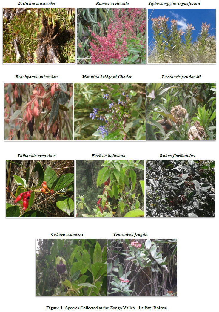
The collected plants belong to eleven different species and families. The species were collected at one of the following altitudinal stages in the valley: High Andean prairie (from 4200 to 4800 m.a.s.l.), Yungas’ Tundra (from 3600 to 4200 m.a.s.l.), Yungas mountain brow (from 2800 to 3600 m.a.s.l.) and Yungas (from 800 to 2800 m.a.s.l.) There is only one specimen belonging to the High Andean prairie, two from the Yungas’ Tundra, six found in the Yungas mountain brow and two appertain to the Yungas’ region. All collected species present colorful organisms (flowers, fruits, leaves or aerial body).
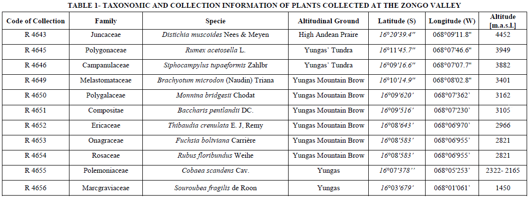
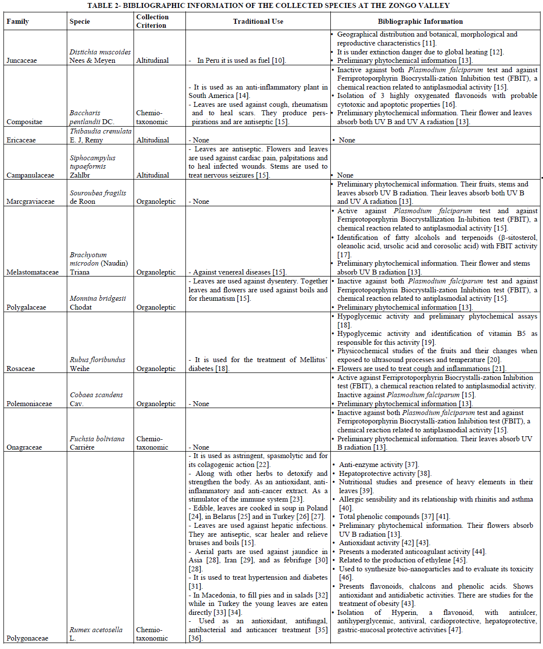
The eleven plants collected are shown in Figure 1 while Table 1 shows the taxonomic information (family and specie) and the data acquired at the collection site (altitudinal ground, coordinates and altitude) for each specie. Table 2 presents a summary of the bibliographic information of each of the collected specie as well as their traditional uses.
3.2 Extract preparation
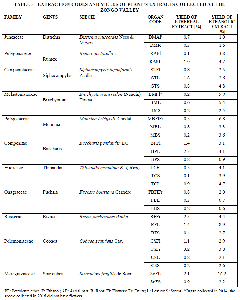
A total of sixty-two vegetal extracts were obtained, thirty-one from the ethereal extraction and thirty-one with the ethanolic procedure. Table 3 shows the summary of the extraction codes and the yield of each organ’s extract. It is important to highlight that for the antioxidant assays only the ethanolic extract were tested since the ethereal extracts were insoluble in the DPPH media. In addition, the tested extracts of Brachyotum microdon flowers where those obtained in a previous collection (2014) since in the collection trip for this publication (2016) we were unable to find this plant with inflorescence.
3.3 DPPH antioxidant assay
The ethanolic extracts were evaluated with the DPPH test at 3 concentrations: 100, 10 and 1µg/ml. Table # 4 shows the results of these antioxidant tests. As it can be seen, most of the extracts present antioxidant activity at 100µg/ml; however, the selection of interesting samples must be done at lower concentrations as compared with the control (ascorbic acid). Therefore, the extracts with interesting results (with activities close to the control at 10µg/ml) are: Baccharis pentlandii (flowers with 91.3% of free radicals trapping capacity), Rubus floribundus (fruits, 90.2%), Fuchsia boliviana (flowers and fruits, 86.7%), Brachyotum microdon (flowers, 82.5%) and Thibaudia crenulate (leaves, 82.1%). Nevertheless, Fuchsia boliviana (leaves) present a higher activity (93.2%) compared to the control (ascorbic acid with 90.0%). These species could have their minimum free radical trapping concentration between 10 and 1µg/ml presenting promising antioxidant activities. In addition, Rumex acetosella, Thibaudia crenulate and Fuchsia boliviana present good antioxidant activities in various organs. Among these species, the genus Baccharis has reported important antioxidant activities which validates our assays and presents this genus as a natural control for antioxidant tests [48], [49], [50], [51], [52], [53].
3.4 Spectroscopic studies
It has been reported that HPLC and spectrophotometric techniques (UV-Vis) are applied to monitor assays used to analyze antioxidant capacities [4]. Based on this work, we have studied the absorption potential of the collected plants and their photoprotective activities in the regions of 280 to 320nm (UV-B) and between 320 to 400nm (UV-A). These spectroscopic studies were carried out using a UV-VIS spectrophotometer and a wavelength window between 200 to 500 nm. Table # 5 presents the samples that have important absorptions in the UV-A and UV-B regions. Some samples were run at a smaller window than 200 to 500 nm and the new window is specified in Table #5. For exemplification purposes, the UV-B absorptions are presented in blue while the UV-A ones are in red.
a. UV analysis for ethereal extracts
The 31 ethereal extracts were studied at 100 ppm in petroleum ether-methylene chloride solvent mixtures. As shown in Table # 5, the species that present important UV-B absorptions are Rumex acetosella (flowers and stems), Brachyotum microdon (leaves and stems), Monnina bridgessi (flowers/fruits), Fuchsia boliviana (flowers/fruits), Thibaudia crenulata (leaves) and Rubus floribundus (fruits).
b. UV analysis for ethanolic extracts
The 31 ethanolic extracts were studied at 100 ppm, some of the samples were also tested at 50 and 25 ppm depending on the plant’s collected amount. As shown in Table # 5, it is important to highlight Fuchsia boliviana (flowers/fruits) absorbing the UV-A and UV-B radiations. Other important species are Baccharis pentlandii and Thibaudia crenulata whose flowers absorb the dangerous UV-A radiation. The species that presents interesting UV-B absorptions are: Brachyotum microdon (flowers), Monnina bridgessi (flowers/fruits), Siphocampylus tupaeformis (flowers and leaves), Fuchsia boliviana (leaves), Souroubea fragilis (stems), Cobaea scandens (flowers, fruits, leaves and stems) and Rubus floribundus (fruits, leaves and stems).
3.5 Analysis of global results (Antioxidant activity and Spectroscopic data)
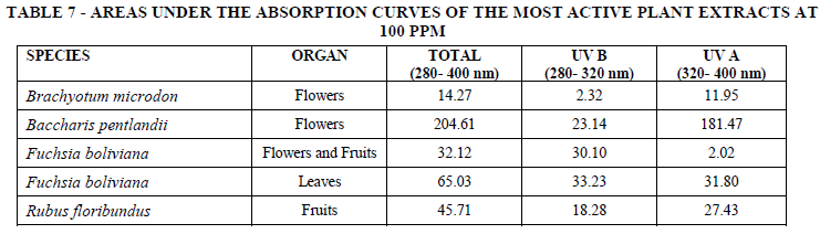
For comparative purposes, Table #6 presents a summary of the global results of the samples with antioxidant activity and their corresponding UV absorptions. As can be seen in this table, there is a strong correlation between the antioxidant activity of a sample and its UV absorptions. Samples at 10µg/ml with free radical trapping capacities above 83% present intense UV- A or/and UV- B absorptions. In Table #6 and for exemplification purposes, DPPH activities above 83% are highlighted in green, UV-B absorptions in blue and UV-A absorptions in red. We can highlight the leaves of Fuchsia boliviana and the flowers of Baccharis pentlandii that present the highest DPPH activity and the highest UV-B and UV-A absorptions, respectively. The fruits of Rubus floribundus are also important for having a DPPH activity above 90% and a high abortion (3.000) in the UV-B region. Figures #2 and #3 show the absorption spectra of the active samples. In Figure #2 we can appreciate the spectra of Fuchsia boliviana (leaves), Baccharis pentlandii (flowers), Rubus floribundus (fruits) and Brachyotum microdon (flowers) all run at 100 ppm. Figure #3 presents the UV absorptions of Fuchsia boliviana (flowers and fruits) assessed at 100ppm and 50ppm. This specie also has a very high DPPH value (91%) and strong absorptions in both UV-B and UV-A regions. Finally, Table #7 presents the areas under the absorption curves of the most active plant extracts at 100 ppm.
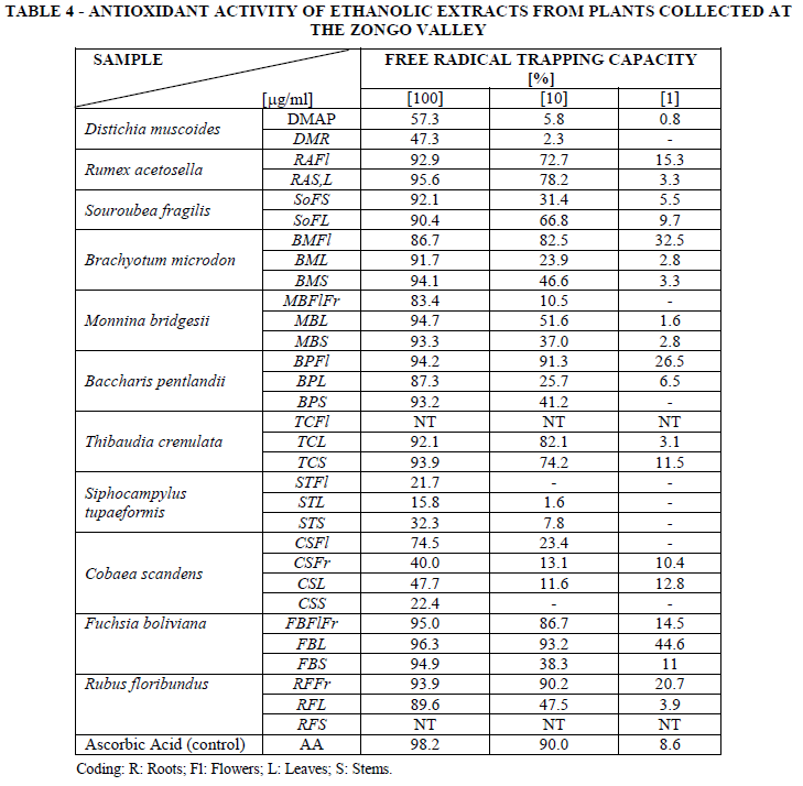
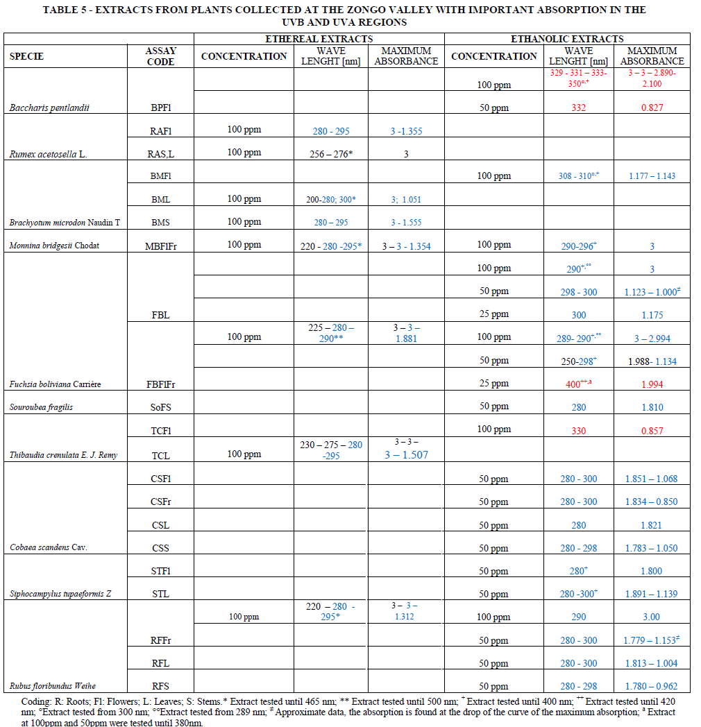
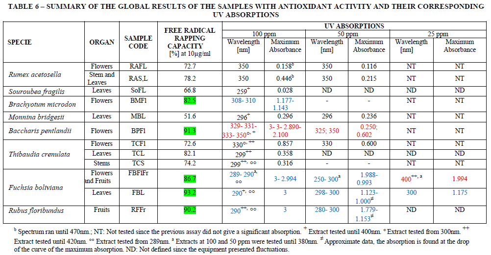
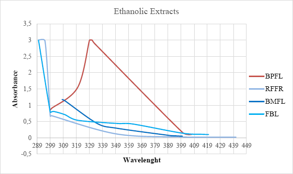
Figure 2: UV spectra of the most active antioxidant samples run at 100ppm.
|
*Spectra run until 350nm. | 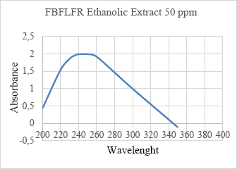 |
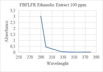
Figure 3: UV spectra of Fuchsia boliviana (flowers and fruits) assessed at 100 and 50ppm*.
4. CONCLUSIONS
Eleven plants were collected at the Zongo valley that provided sixty two vegetal extracts, thirty one from the ethereal extraction and thirty one with the ethanolic procedure. The ethanolic extracts were essayed with the DPPH test in order to find antioxidant samples. In addition, the UV absorptions of all extracts were also evaluated. Twelve extracts showed free radical trapping capacity above 50% when tested at 10µg/ml. The most active samples (activities above 83%) presented strong UV-B (280- 320nnm) and/or UV-A (320- 400nm) absorptions. It is important to highlight the leaves of Fuchsia boliviana with 93.2% in the DPPH assay and with a strong absorption (3.000 A) in the UV-B region (290nm); the flowers of Baccharis pentlandii (91.3% DPPH, 329-331nm/3A); the fruits of Rubus floribundus (90.2% DPPH, 290nmn/3A); the flowers and fruits of Fuchsia boliviana (86.7% DPPH, 289-291nm/3A; 400nm/2A) and the flowers of Brachyotum microdon (82.5% DPPH, 308nm/1.2A). The ultraviolet absorptions of many of the species submitted in this paper were previously reported [13]. Comparing the results from both publications we can define the best season to collect the important species. For instance, Brachyotum microdon (flowers) should be collected in October (spring) while Baccharis pentlandii and Fuchsia boliviana in May (fall). Plants collected in the mentioned season have higher UV-A and/or UV-B absorption. In addition, all the active species present phenolic compounds, flavonoids, flavones and tannins in their composition [13]. The antioxidant activities of these types of compounds were previously presented in the work of Pisoschi et. al. [4]. Moreover, Brachyotum microdon (flowers) also presents anthraquinones, anthocyanins, chalcones and quinones while Baccharis pentlandii (flowers) and Fuchsia boliviana (flowers and fruits) have chalcones and quinones. Finally, Fucshia boliviana (flowers and fruits) also has anthraquinones and isoflavones [13]. These compounds could have biological activities since their molecular structures are close to the active flavonoids and their biosynthetic pathways are those of phenolic compounds (derived from shikimate) [54].
Most of the plants have antioxidant compounds as their main defense mechanisms; therefore, it is not surprising to find many positive results in antioxidant evaluations in vitro [6]; however, if the samples are evaluated at low concentrations (10 or 1µg/ml) the most powerful antioxidants can be detected. In this paper we report a strong correlation between antioxidant activity evaluated by DPPH and UV absorptions. This DPPH/UV technique could be used to preliminary screen vegetable samples and select those with DPPH values above 83% and strong UV-A or UV-B absorptions. The chosen samples, then can be evaluated with other more expensive in vitro assay (TEAC, ABTS or FRAP) to finally confirm their activities with the in vivo tests.
5. REFERENCES
[1] R. Szymanska, P. Pospisil and J. Kruk, "Plant-Derived Antioxidants in Disease Prevention," Oxidative Medicine and Cellular Logevity , no. Doi: 10.1155/2018/2068370, 2018.
[2] P. Rajendrana, N. Nandakumar, T. Rengarajan, R. Palaniswamid, E. N. Gnanadhas, U. Lakshminarasaiah, J. Gopasb and I. Nishigakia, "Antioxidant and Human Diseases," Clinica Chimica Acta, vol. 436, pp. 332-347, 2014.
[3] K. Rahman, "Studies on free radicals, antioxidants, and cofactors," Clin Interv Aging, , vol. 2, p. 219–236, 2007.
[4] A. M. Pisoschi, A. Pop, C. Cimpeanu and G. Predoi, "Antioxidant Capacity Determination in Plants and Plant-Derived Products: A Review," Oxid Med Cell Longev, no. Doi: 10.1155/2016/9130976., 2016.
[5] E. A. Shalaby and S. M. M. Shanab, "Antioxidant Compounds, Assays of Determination and Mode of Action," African Journal of Pharmacy and Pharmacology, vol. 7, pp. 528-539, 2013.
[6] D. M. Kasote, S. S. Katyare, M. V. Hegde and H. Bae, "Significance of Antioxidant Potential of Plants and its Relevance to Therapeutic Applications," Int. J. Biol. Sci., vol. 11, pp. 982- 991, 2015.
[7] T. Ortuño, Estudio palinológico en un gradiente altitudinal en el valle de Zongo. Tesis de licenciatura en Biología, La Paz- Bolivia: UMSA, 2000.
[8] M. Baudoin, Historia natural de un valle de los Andes, La Paz: Instituto de Ecología-UMSA, 1991. [ Links ]
[9] M. Joyeaux, A. Lobstein, R. Anton and F. Mortier, "Comparative antilipoperoxidant, antinecrotic and scavenging properties of terpenes and biflavones from Ginkgo and some flavonoids," Planta Med. , vol. 61, pp. 126-129, 1995.
[10] H. Balslev, Flowering Plant. Monocotyledons, Springer, 1998. [ Links ]
[11] P. Gonzáles, M. Suni, R. Deanna, M. A. Scaldaferro, E. Castañeda, D. W. Ramirez, N. Valencia and A. Cano, "Reproductive biology and cytogenetics of Distichia muscoides (Juncaceae)," Boletín de la Sociedad Argentina de Botánica, vol. 51, pp. 123-133, 2016.
[12] S. Loza Herrera, R. I. Meneses and F. Anthelme, "Comunidades vegetales de los bofedales de la Cordillera Real (Bolivia) bajo el calentamiento global / Plant communities of high-Andean wetlands of the Cordillera Real (Bolivia) in the face of global warming," Ecología en Bolivia, vol. 50, pp. 39-56, 2015.
[13] S. L. Ibáñez-Calero, K. E. Loayza, E. L. Yapu, J. Lizarazu, R. Zeballos and T. Solares, "A Screening of Natural Colorants with probable antioxidant and/or photo-protector activities," Investigacion y Desarrollo, vol. 16, pp. 5-24, 2016.
[14] M. Abad, A. Bessa, B. Ballarin, O. Aragón, E. Gonzales and P. Bermejo, "Anti-inflammatory activity of four Bolivian Baccharis species (Compositae)," J. Ethnopharmacol., vol. 103, pp. 338-344, 2006.
[15] S. L. Ibáñez-Calero, Application du test d’inhibition de la biocristallisation de l´heme a la caracterisation de composes antipaludiques isoles de la flore de Bolivie. Ph.D. thesis, Toulouse- France: Universite Paul Sabatier, 2005.
[16] S. Tarqui, Y. Flores and G. Almanza, "Polyoxygenated flavonoids from Baccharis pentlandii," Revista Boliviana de Química, vol. 29, pp. 10- 14, 2012.
[17] S. L. Ibáñez-Calero, V. Jullian, S. C. d. M. L. R. Chevalley, J. Bravo, A. Gimenez and M. Sauvain, "Application of Ferriprotoporphyrin Biocrystallization Inhibition Test to Find Antiplasmodial Compounds in the Flora of the Zongo Valley- Bolivia," Revista Boliviana de Química, vol. 29, pp. 71-79, 2012.
[18] F. R. Pérez Azahuanche, J. C. Guerrero Hurtado, Z. M. Ortiz Rubio, F. Rodríguez Ávalos and G. León Aponte, "Análisis fitoquímico preliminar y evaluación de la actividad hipoglucemiante de Rubus loribundus Kunth (Rosaceae) "zarzamora"," Arnaldoa, vol. 21, pp. 391- 401, 2014.
[19] J. C. Guerrero Hurtado, G. León Aponte, F. R. Pérez Azahuanche, F. Rodríguez Ávalos and Z. M. Ortiz Rubio, "Aislamiento y caracterización estructural de los principios activos hipoglucemiantes de Rubus floribundus Kunth (Rosaceae) “zarzamora"," Arnaldoa, vol. 22, pp. 381- 394, 2015.
[20] C. V. Pérez Rodríguez and F. R. Pérez Azahuanche, "La temperatura en las propiedades fisicoquímicas, contenido de vitamina C y recuento de mohos y levaduras del mesocarpio de “zarzamora” Rubus floribundus kunth (Rosaceae)," Arnaldoa, vol. 26, pp. 297-304, 2019.
[21] I. H. Castillo Vera, Importancia cultural de la flora silvestre utilizada por los pobladores del caserío de Cabrero en la microcuenca Quebrada Honda (Cajabamba, Cajamarca, Perú) TESIS Para optar el Grado Académico de Magíster en Botánica Tropical con mención en Etnobotánica, Lima-Peru: Universidad Nacional Mayor de San Marcos Universidad del Perú, 2018.
[22] M. Wegiera, H. Smolarz and Bogucka-Kocka, "Rumex L. species induce apoptosis in 1301, EOL-1 and H-9 cell lines," A. Acta Pol Pharm., vol. 69, pp. 487-499, 2012.
[23] N. C. Institute, "PDQ Cancer Information Summaries [Internet]. US. February 3, 2014.," US, USA, 2014. [ Links ]
[24] Ł. Łuczaj and W. Szymański, "Wild vascular plants gathered for consumption in the Polish countryside: a review," J Ethnobiol Ethnomed. , vol. 15, pp. 3-17, 2007.
[25] Ł. Łuczaj, P. Köhler, E. Pirożnikow, M. Graniszewska, A. Pieroni and T. Gervasi, "Wild edible plants of Belarus: from Rostafiński’s questionnaire of 1883 to the present," J Ethnobiol Ethnomed, no. doi: 10.1186/1746-4269-9-21, 2013.
[26] S. Kültür, "An ethnobotanical study of Kırklareli (Turkey)," Phytologia Balcanica, vol. 14, p. 279 –289, 2008.
[27] T. Özen, "Antioxidant activity of wild edible plants in the Black Sea Region of Turkey," Grasas y aceites, no. search.ebscohost.com. , 2010.
[28] M. S. Amiri, M. R. Joharchi and M. E. TaghavizadehYazdi, "Ethno-Medicinal Plants Used to Cure Jaundice by Traditional Healers of Mashhad, Iran," Iranian Journal of Pharmaceutical Research, vol. 13, pp. 157-162, 2014.
[29] D. Tewari, A. Mocan, E. D. Parvanov, A. N. Sah, S. M. Nabavi, L. Huminiecki, Z. F. Ma, Y. Y. Lee, J. O. Horbańczuk and A. G. Atanasov, "Ethnopharmacological Approaches for Therapy of Jaundice: Part I," Front Pharmacol., no. doi: 10.3389/fphar.2017.0051, 2017.
[30] M. S. Amiri, M. R. Joharchi and M. E. TaghavizadehYazdi, "Ethnobotanical investigation of traditional medicinal plants commercialized in the markets of Mashhad, Iran," Avicenna. J Phytomed., vol. 3, no. 3, p. 254–271, 2013.
[31] W. Kipkore, B. Wanjohi, H. Rono and G. Kigen, "A study of the medicinal plants used by the Marakwet Community in Kenya," J. Ethnobiol. Ethnomed., no. doi: 10.1186/1746-4269-10-24, 2014.
[32] A. Pieroni, B. Rexhepi, A. Nedelcheva, A. Hajdari, B. Mustafa, V. Kolosova, K. Cianfaglione and C. L. Quave, "One century later: the folk botanical knowledge of the last remaining Albanians of the upper Reka Valley, Mount Korab, Western Macedonia," J. Ethnobiol. Ethnomed., no. doi: 10.1186/1746-4269-9-22, 2013.
[33] S. Arı, M. Temel, M. Kargıoğlu and M. Konuk, "Ethnobotanical survey of plants used in Afyonkarahisar-Turkey," J. Ethnobiol. Ethnomed., no. doi: 10.1186/s13002-015-0067-6, 2015.
[34] G. Tuttu, G. Abay and S. Yildirimli, "Some Wild Edible Plants of Tosya District (Kastamonu, Turkey)," International Journal of Scientific and Technological Research, vol. 5, no. 3, pp. 129-135, 2019.
[35] A. Vasas, O. Orbán-Gyapai and J. Hohmann, "The genus Rumex: review of traditional uses, phytochemistry and pharmacology," J. Ethnopharmacol., vol. 175, pp. 198- 228, 2015.
[36] C. Lans, "Do recent research studies validate the medicinal plants used in British Columbia, Canada for pet diseases and wild animals taken into temporary care?," Journal of Ethnopharmacology, vol. 236, pp. 366-392, 2019.
[37] D. Ahmed, Q. Mughal, S. Younas and M. Ikram, "Study of phenolic content and urease and alpha-amylase inhibitory activities of methanolic extract of Rumex acetosella roots and its sub-fractions in different solvents," Pak. J. Pharm. Sci., vol. 26, pp. 553-559, 2013, 2013.
[38] N. Zarrin Sarhady, B. Ali Mandegary, S. Fariba, M. Saeedeh and I. Maryam, "Evaluation of the hepatoprotective effects of Rumex acetosella on the carbon tetrachloride-induced hepatotoxicity in rats," Avicenna Journal of Phytomedicine, vol. 5, pp. 134-135, 2015.
[39] S. Fatma Zerrin and C. Hale Seçilmiş, "Determination of Heavy Metals and Nutrient Elements in Some Medicinal Plants Used in Eskişehir," Journal of Natural & Applied Sciences, vol. 19, pp. 83-90, 2015.
[40] O. Rodríguez, R. Célio, F. Aboukhair, A. M. Laurrabaquio, I. O. Tinoco, H. U. Cuevas, M. A. Cruz Suárez, M. A. Cruz Marmolejo and M. C. Reyes, "Prueba cutánea con extractos alergénicos de pólenes y relación con signos clínicos de rinitis alérgica y asma bronquial en Camagüey, Cuba," Revista Vacci Monitor (Vacunología y Temas Afines), vol. 22, pp. 9-13, 2013.
[41] L. Tejeda, Antioxidants in Andean food and meals. PhD Thesis in Food Engineering, Lund: Lund University Publications, 2013.
[42] B. Zengin Kurt, I. Gazioğlu, E. Sevgi and F. Sönmez, "Anticholinesterase, Antioxidant, Antiaflatoxigenic Activities of Ten Edible Wild Plants from Ordu Area, Turkey, Iran," J Pharm Res., vol. 17, no. 3, p. 1047–1056, 2018.
[43] S. Sekhon-Loodu and H. P. Rupasinghe, "Evaluation of antioxidant, antidiabetic and antiobesity potential of selected traditional medicinal plants," Frontiers in nutrition, no. doi.org/10.3389/fnut.2019.00053, 2019.
[44] M. Alikhani Pour, S. Sardari, A. Eslamifar, A. Azhar, M. Rezvani and M. Nazari, "Cheminformatics-Based Anticoagulant Study of Traditionally Used Medicinal Plants," Iran Biomed. J. , vol. 21, no. 6, p. 400–405, 2017.
[45] M. Banga, E. J. Slaa, C. W. P. M. Blom and L. A. C. J. Voesenek, "Ethylene Biosynthesis and Accumulation under Drained and Submerged Conditions (A Comparative Study of Two Rumex Species)," Plant Physiol. , vol. 112, no. 1, p. 229–237, 1996.
[46] I. Khan, A. Bahuguna, M. Krishnan, S. Shukla, H. Lee, S. Min, D. Kyu Choi, Y. Cho, V. K. Bajpai, Y. Suk Huh and S. Chul Kang, "The effect of biogenic manufactured silver nanoparticles on human endothelial cells and zebrafish model," Science of the Total Environment, vol. 679, p. 365–377, 2019.
[47] N. Verma, A. Pratap Singh and C. V. Rao, "Antioxidant mediated ulcer healing potential of hyperin isolated from flowers of rhododendron arboreum in experimental rat," International journal of pharmaceutical sciences and research, vol. 10, no. 3, pp. 1524- 1533, 2019.
[48] S. Q. De Oliveira, F. Dal-Pizzol, J. C. F. Moreira, E. P. Schenkel and G. Gosmann, "Antioxidant activity of Baccharis spicata, Baccharis trimera and Baccharis usterii," Acta Farm. Bonaerense, vol. 23, no. 3, pp. 365-368, 2004.
[49] C. B. De Oliveira, L. N. Comunello, A. Lunardelli, R. H. Amaral, M. G. S. Pires, G. L. Da Silva, V. Manfredini, C. Regla Vargas, S. C. B. Gnoatto, J. R. De Oliveira and G. Gosmann, "Phenolic Enriched Extract of Baccharis trimera Presents Anti-inflammatory and Antioxidant Activities," Molecules, vol. 17, no. 1, pp. 1113-1123, 2012.
[50] S. Q. De Oliveira, F. Dal-Pizzol, G. Gosmann, D. Guillaume, J. C. F. Moreira and E. P. Schenkel, "Antioxidant Activity of Baccharis articulata Extracts: Isolation of a New Compound with Antioxidant Activity," Free Radical Research, vol. 37, no. 5, pp. 555-559, 2003.
[51] B. Da Cruz Pádua, L. D. Silva, J. V. Rossoni Júnior, J. L. Humberto, M. Martins Chaves, M. E. Silva, M. L. Pedrosa and D. Caldeira Costa, "Antioxidant properties of Baccharis trimerain the neutrophils of Fisher rats," Journal of Ethnopharmacology, vol. 129, no. 3, pp. 381-386, 2010.
[52] M. J. Simirgiotis, C. Quispe, J. Bórquez, A. Mocan and B. Sepúlveda, "High resolution metabolite fingerprinting of the resin of Baccharis tola Phil. from the Atacama Desert and its antioxidant capacities," Industrial Crops and Products, vol. 94, pp. 368-375, 2016.
[53] S. M. Sabir, M. L. Athayde, A. A. Boligon and J. B. T. Rocha, "Antioxidant activities and phenolic profile of Baccharis trimera, a commonly used medicinal plant from Brazil," South African Journal of Botany, vol. 113, pp. Pages 318-323, 2017.
[54] J. Mann, Secondary Metabolism, 2nd edition, Oxford: Oxford Science Publications, 1992.

