


 texto en
  Inglés (pdf)
 Articulo en XML
 Referencias del artículo
 Enviar articulo por email
 Citado por SciELO
  Similares en
SciELO
Similares en
SciELO  uBio
uBio 
 Permalink
Journal of the Selva Andina Animal Science
versión impresa ISSN 2311-3766versión On-line ISSN 2311-2581
J.Selva Andina Anim. Sci. v.8 n.1 La Paz 2021
https://doi.org/10.36610/j.jsaas.2021.080100003x
Short Communication
Caracterization ultrasonographic B-mode and Doppler of the corpus luteum in llamas
Caracterización ultrasonográfica modo-B y Doppler del cuerpo lúteo en llamas
Perez Guerra Uri Harold1*![]() , Bustamante Quispe Carlos Washington1
, Bustamante Quispe Carlos Washington1![]() , Luque Mamani Natalio2
, Luque Mamani Natalio2![]() , Huayta Arizaca Rito Felipe3
, Huayta Arizaca Rito Felipe3![]() , Condori Chuchi Eloy Amador2
, Condori Chuchi Eloy Amador2![]() , Catacora Flores Nubia Lilia4
, Catacora Flores Nubia Lilia4![]() , Pérez Durand Manuel Guido1
, Pérez Durand Manuel Guido1![]()
1National University of the Altiplano Puno. Faculty of Veterinary Medicine and Zootechnics. Laboratory of Animal Reproduction. Panama Avenue
No 710. Tel: +051-599430. Puno, Peru.
2National
University of the Altiplano Puno. Faculty
of Veterinary Medicine and Zootechnics. Chuquibambilla
Research and Production Center. Animal
Health Laboratory. Umachiri
District, Melgar Province. Puno, Peru. Tel:
+051-599430.
3National
University of the Altiplano Puno. Postgraduate School Doctorate
in Animal Science. Av.
Floral Nº 1153. Tel: + 051-599430. Puno,
Peru.
4Universidad
Nacional del Altiplano Puno. Faculty
of Veterinary Medicine and Zootechnics. Chuquibambilla
Research and Production Center. Laboratory
of Reproductive Biotechnology. District
of Umachiri, Province of Melgar. Tel:
+ 051-599430. Puno, Peru.
*Contact address: National University of the Altiplano Puno. Faculty of Veterinary Medicine and Zootechnics. Laboratory of Animal Reproduction. Panama Avenue No 710. Tel: +051-599430. Puno, Peru.
Uri Harold Perez Guerra
E-mail address : uperez@unap.edu.pe
Record from the
article
Received September
2020.
Returned November
2020.
Accepted January
2021.
Available online,
April 2021.
ID of article: 083/JSAAS/2020
J. Selva Andina Anim. Sci. 2021; 8(1):3-11.
Abstract
B-mode and Doppler ultrasonography in ruminants, as a technique has allowed establishing new concepts on the reproductive physiology of females, through the study of follicular dynamics and morphometry of the corpus luteum, therefore, the objective was to characterize by means of B-mode and Doppler ultrasonography the corpus luteum in recipient llamas. Thirty-seven recipient llamas were used (27 for B-mode ultrasonography and 10 Doppler) that were synchronized by applying buserelin acetate 0.0096 mg, nine days later 0.048 mg of prostaglandin F2α analog was applied two days later, the second dose of buserelin acetate was applied at the same dose to guarantee ovulation and subsequent formation of the corpus luteum, the evaluation in mode B of the morphometry consisted of observing the echotexture, area, diameter and volume of the corpus luteum with the use of a SonoStar SS-8® ultrasonograph at 6.5 MHz frequency and 6 cm depth equipped with a transrectal linear transducer; using the same technique with a Draminski 4Vet® Doppler equipment, the percentage of luteal area of vascularization (% AVL) was determined. The data were subjected to a descriptive analysis being 0.119±0.032 cm2 of area, 12.7±1.7 mm of diameter and 0.84±0.32 cm3 of CL volume, to determine the relationship between metric measures the Pearson correlation was used observing a positive relationship between area/diameter of 0.7506, area/volume of 0.9289 and diameter/volume of 0.6602, observing a high positive correlation between area and volume, finally the % AVL characteristics was 34.97 for the CL of recipient llamas. In conclusion, the morphometric characteristics evaluated in mode B (area, diameter and volume) have a positive correlation and the % AVL could be applied as a tool in the efficient reproductive management for the selection of recipients in camelids, understanding that it is one of the first reports of these characteristics in llamas.
Keywords: Corpus luteum, Doppler, llama, morphometry, pregnancy, ultrasonography.
Resumen
La ultrasonografía modo B y Doppler en los rumiantes, como técnica ha permitido establecer nuevos conceptos sobre la fisiología reproductiva de las hembras, a través del estudio de la dinámica folicular y morfometría del cuerpo lúteo, por tanto, el objetivo fue caracterizar mediante ultrasonografía modo B y Doppler el cuerpo lúteo en llamas receptoras. Se utilizaron 37 llamas como receptoras (27 para ultrasonografía modo B y 10 Doppler) que fueron sincronizadas aplicando acetato de buserelina 0.0096 mg, nueve días posteriores se aplicó 0.048 mg de análogo de prostaglandina F2α, a los dos días posteriores se aplicó la segunda dosis de acetato de buserelina en la misma dosis para garantizar la ovulación y posterior formación del cuerpo lúteo, la evaluación en modo B de la morfometría consistió en observar la ecotextura, área, diámetro y volumen del cuerpo lúteo con el uso de un equipo ultrasonógrafo SonoStar SS-8® a 6.5 MHz de frecuencia y 6 cm de profundidad equipado con un transductor lineal transrectal; con la misma técnica con un equipo Draminski 4Vet® Doppler se determinó el porcentaje de área luteal de vascularización (% AVL). Los datos fueron sometidos a un análisis descriptivo siendo de 0.119±0.032 cm2 de área, 12.7±1.7 mm de diámetro y 0.84±0.32 cm3 de volumen de CL, para determinar la relación entre medidas métricas se utilizó la correlación de Pearson observando una relación positiva entre área/diámetro de 0.7506, área/volumen de 0.9289 y diámetro/volumen de 0.6602, observando una correlación alta positiva entre área y volumen, finalmente las características de % AVL fue de 34.97 para los CL de llamas receptoras. En conclusión, las características morfométricas evaluadas en modo B (área, diámetro y volumen) tienen una correlación positiva y el % AVL podría aplicarse como herramienta en el manejo reproductivo eficiente para la selección de receptoras en camélidos, entendiendo que es uno de los primeros reportes de estas características en llamas.
Palabras clave: Cuerpo lúteo, Doppler, llama, morfometría, preñez, ultrasonografía.
Introduction
Efficient reproductive management and the use of reproductive biotechnologies such as artificial insemination (AI) and embryo transfer (ET) require an understanding of the follicular dynamics that govern ovarian activity1. Several studies exist on follicular dynamics in South American camelids (SAC) characterizing a continuous and overlapping wave with induced ovulation2-4. The detection of corpus luteum (CL) by rectal palpation by experts has a positive predictive value of only 64 %, even confusing between follicles and CL, while ultrasonography (US) allows the identification of CL with an efficiency of 85 %5,6.
The wide availability of the US has facilitated the continuous advancement of knowledge of the physiological changes and characteristics of the CL during the estrous cycle in cattle7. The CL, through the production of progesterone (P4), plays a key role in the establishment and, maintenance of pregnancy in all domestic animals8, including camelids, especially in ET programs for the selection of recipient females9-11. US allows the evaluation of ovarian morphometric characteristics such as follicles, CL, among others. Studies with US evaluating the CL make it possible to determine the reproductive status in cattle, monitor its dynamics, formation, development, and regression of this structure12. The ultrasound image of the LC is uniform, circumscribed and less echogenic than the ovarian stroma, the sound wave intensity. The return of the echogenicity and echotexture, characteristics that are used as a potential measure for predicting LC function and steroidogenic capacity13,14 can be used for the evaluation of echogenicity and echotexture. Physiological and morphometric studies in reproduction evaluated with US Doppler of the LC in humans report a positive correlation between the LC blood perfusion index and progesterone concentrations in early pregnancies, so in cattle and camelids it could also predict early pregnancy, opening new knowledge even more if factors such as altitude are included on the physiology of domestic animals in these environmental conditions, assuming slight variations compared with studies carried out at other altitudes15-17, for this reason it is necessary to use US in the study of reproductive physiology in domestic animals that inhabit the Peruvian highlands, for this reason the objective of the present study was to characterize the corpus luteum in recipient llamas by means of B-mode ultrasonography and Doppler.
Materials and methods
Animals. A total of 37 recipient llamas were selected from an embryo transfer program for B-mode ultrasonographic evaluation and 10 of them for Doppler evaluation (due to the complexity and time- consuming nature of the Doppler study), the study was carried out at the Research and Production Center (RPC) "La Raya" of the National University of the Altiplano Puno (UNAP), located at 4230 meters above sea level, where the llamas had at least one calving, more than 15 days post parturition (empty llamas), with a corporal condition of 3.44 on a scale of 1 to 518, fed with natural pasture.
Wave synchronization and ovulation induction in recipients. For synchronization, GnRH (buserelin acetate: 0.0096 mg: Gestar® - Over - Argentina) was applied on day "0" and a prostaglandin F2α analogue (PGF: 0.048 mg: Cloprostenol: Prostal® Over - Argentina), two days later (day 11) a second dose of GnRH analog (buserelin acetate: 0.0096 mg: Gestar® - Over - Argentina) was applied to ensure ovulation and formation of the CL, finally the ultrasonographic evaluation was performed on the 18th day of treatment19.
Ultrasonographic evaluation of CL morphometry. The evaluation of the morphometry in mode B consisted of taking characteristics such as: area, volume, and diameter with a SonoStar SS-8® (SonoStar Technologies, China) ultrasonographic equipment with 6.5 MHz frequency and 6 cm depth equipped with a linear transrectal transducer, the CL was located by means of the US equipment (SonoStar SS-8®) in mode 2B to capture two images through its freeze option and evaluate its characteristics as described below: i) Volume of the LC. Three diameters were taken over the CL by placing the calipers (electronic calipers of the US software) in the limit between the luteal tissue and the ovarian stroma to be valuated and immediately the veterinary software ofthe US equipment (SonoStar SS-8®) allowed us to visualize the volume of the CL in cm3 (Figure 1). ii) Area of the CL. The option of circular area was chosen due to the characteristics of the CL, the calipers were placed in the limit between the luteal tissue and the ovarian stroma. This circular area was then opened, which allowed us to obtain the area of the CL in cm2. iii) CL diameter. calipers were placed between the luteal tissue and the visualized ovarian stroma, which allowed us to obtain the diameter of the CL in mm.
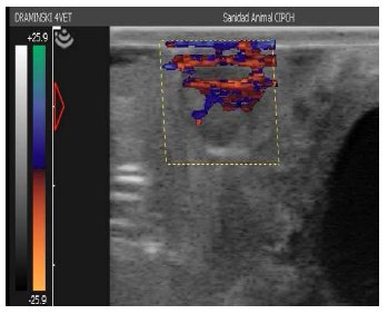
The evaluation of the Doppler ultrasonographic morphometry consisted of determining the area of luteal vascularization (AVL %) with a US Draminski 4Vet® (Draminski ul. Owocowa 17, Poland) with 6.5 MHz frequency and 6 cm depth equipped with a linear transrectal transducer, the CL was placed in B mode, then the Color Doppler mode was activated to determine the vascular activity of the CL, the images were stored and analyzed in the Image J® program, determining the AVL in percentage using the following formula20:
![]()
Statistical analysis. The B-mode and Doppler morphometric characteristics, being a characterization study and one of the first reports, were determined by descriptive statistics (average and standard deviation) of volume, area, diameter and AVL (%), specifically the B-mode characteristics were subjected to Pear's correlation to determine their degree of association; all data were processed using the statistical program R version 3.5.1.21.
Results
The echotexture of CL in llamas is hypoechogenic (darker) compared to the ovarian stroma due to its greater vascularization. The most important characteristic of the CL in llamas is that it is not embedded in the ovary but appears to be an appendage of the ovary. Table 1 shows the metric characteristics (area, diameter and volume) of the CLs evaluated by US mode B in llamas.
Table 1 Morphometric characteristics of CL (area, diameter and volume: average and standard deviation, respectively) of B-mode llamas |
|
In all the characteristics evaluated, a positive correlation was observed between area/diameter of 0.7506, area/volume of 0.9289, and diameter of 0.9289. The significant association between area and volume of the CL would allow taking only one of these characteristics in subsequent ultrasonographic evaluations of CL in llamas, the coefficients of variation of the evaluated characteristics were for área of 26.9 %, for diameter 13.4 % and volume of 38.1 %, observing less variability in the determination of the diameter of CL.9 %, for diameter 13.4 % and volume of 38.1 % observing a lower variability in the determination of the diameter of CL, the morphometric characteristics by US are as shown in figure 1.
Figure 1 Ultrasonographic evaluation of the morphometric characteristics and
echotexture of the CL of recipient llamas in B-mode |
|
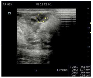
Figure 2 shows the AVL determined as a percentage of the proportion of blood flow that irrigates the CL at the time of the evaluation (7 days after the application of GnRH), with an average of 34.97% of the total área of the LC evaluated, it should be noted that the red color of the image corresponds to the arterial vascularization while the blue color corresponds to the venous vascularization, which could be specified in subsequent studies.
Figure 2 Ultrasonographic evaluation of the Doppler characteristics of the CL of recipient llamas |
|
Discussion
The echotexture of the LC of recipient llamas effectively shows them to be uniform, circumscribed, hypoechogenic and homogeneous structures (Figure 1), due to the fact that their ovarian stroma is related to the active angiogenic process that occurs during the initial stages of luteal development in mammals, mediated by angiogenic factors such as vascular endothelial growth factor (VEGF), whose increase leads to rapid mitosis of the endothelial cells of the capillaries of the LC1422,23, in addition to the fact that most steroidogenic cells are adjacent to one or more capillaries and the perfusion of the luteal blood flow is so intense that it affects the acoustic impedance of the organ, resulting in the characteristic echotexture of the LC1224, as occurs in other species with the same hypoechogenic characteristics in relation to the ovarian stroma25,26.
In B-mode ultrasonography of the CL in recipient llamas show morphometric characteristics such as area, volume and diameter highly related to studies on follicular dynamics in llamas specifically with sizes of donor follicles reaching diameters of 10.0±2.0 mm and 11.8±1.6 mm respectively3,27, the maximum follicular diameter is related to the future diameter of the CL since these follicles have active angiogenesis after theca formation, which promotes the permeability of blood vessels, ovulation and subsequent formation of the CL28, in cows for meat and milk production, ovulation of a preovulatory follicle of greater diameter leads to a CL of good size; this characteristic seems to be associated with fertility29,30. The diameter of the CL of the llamas was 12.7±1.7 mm, compared to other studies, are similar to those reported at 7 days post hormone application reaching diameters of 12.3±0.6 mm, 10.4±0.4 mm, 11±1.9, 11.19±0.32 mm31-33. While the area of the CL was 0.119±0.03 cm2 and the volume of the CL evaluated was 0.84±0.34 cm3, these being the first results reported with various morphometric measurements of the CL and their correlation between them in llamas by US.
In ultrasonographic Doppler morphometry of the CL in recipient llamas and specifically Color Doppler allows to determine the blood flow denoted as AVL in percentage, because the CL is highly vascularized and shows higher blood flow per unit of body tissue, the CL is characterized by intense angiogenesis and the CL blood flow increases 3 to 4 times in the first 96 h after ovulation in cattle20, in the present study it was determined that the blood flow represented by the AVL was 34.In contrast, in llamas, the AVL between day 6 and 8 post application of GnRH was 20 and 25 %34, while other authors report between 30 and 40 % of AVL, similar results to the present study35, which is why similar percentages of AVL are determined in high altitude conditions, This evaluation performed with color Doppler allows the evaluation of the quantity and pattern of blood flow in the LC, which indirectly indicates its functionality, a useful tool for reproductive management decisions such as the objective selection of recipients in embryo transfer programs in camelids, as well as mainly in cattle36,37. In conclusion, the morphometric characteristics evaluated in B-mode (area, diameter and volume) have a positive correlation, therefore, any of the metric characteristics of the CL could be used for the selection of recipients in camelids, and the AVL percentage could be applied as a predictive tool for efficient reproductive management, diagnosis of early pregnancy and for the selection of recipients in camelids, as occurs in cattle.
Funding source
This article has not received any funding other than from the research group's own resources.
Conflicts of interest
The authors declare that the present research was carried out at the National University of the Altiplano Puno and there is no conflict of interest between the authors of this article.
Acknowledgments
To the Animal Health Laboratory of the Chuquibambilla Research Center for the loan of the Doppler ultrasound equipment, as well as to the Experimental Center of "La Raya" for the use of the experimental material (llamas under study).
Ethical considerations
The study was approved by the Ethics Committee of the Faculty of Veterinary Medicine and Zootechnics of the National University of the Altiplano Puno and the guidelines established by this Committee were followed.
Authors' contribution to the article
Perez Guerra Uri Harold performed the experimental design and drafting of the manuscript, Bustamante Quispe Carlos Washington developed the experimental part of the ultrasonographic B-mode, Luque Mamani Natalio reviewed and wrote the manuscript, Huayta Arizaca Rito developed the experimental part of the ultrasonographic Doppler, Condori Chuchi Eloy Amador design of the experiment and statistical analysis, Catacora Flores Nubia Lilia performed the statistical analysis and revision of the manuscript, Pérez Durand Manuel Guido performed the writing and final revision of the manuscript.
Cited Literature
1. Tibary A. Monitoring and controlling follicular activity in camelids. Theriogenology 2018;109: 22-30. DOI: https://doi.org/10.1016/j.theriogenology.2017.12.011
2. Cavilla MV, Bianchi CP, Maistruarena C, Aba MA. Ultrasonographic and endocrine characterization of follicular waves in llamas with a special reference to the overlapping phenomenon during successive waves. Reprod Domest Anim 2013;48(6):923-30. DOI: https://doi.org/10.1111/rda.12187
3. Chaves MG, Aba M, Agüero A, Egey J, Berestin V, Rutter B. Ovarian follicular wave pattern and the effect of exogenous progesterone on follicu-lar activity in non-mated llamas. Anim Reprod Sci 2002;69(1-2):37-46. DOI: https://doi.org/10.1016/s0378-4320(01)00173-7
4. Perez U, Pari D, Gutierrez F, Málaga J, Luque N, Rojas R, et al. Comparación ultrasonográfica transvaginal y transrectal de la dinámica folicular en ondas sucesivas de llamas (Lama glama). Rev Investig Vet Perú 2021;32(1):e19504. DOI: https://doi.org/10.15381/rivep.v32i1.19504
5. Herzog K, Brockhan-Lüdemann M, Kaske M, Beindorff N, Paul V, Niemann H, et al. Luteal blood flow is a more appropriate indicator for luteal function during the bovine estrous cycle than luteal size. Theriogenology 2010;73(5):691-7. DOI: https://doi.org/10.1016/j.theriogenology.2009.11.016
6. Ribadu AY, Ward WR, Dobson H. Comparative evaluation of ovarian structures in cattle by pal-pation per rectum, ultrasonography and plasma progesterone concentration. Vet Rec 1994;135 (19):452-7. DOI: https://doi.org/10.1136/vr.135.19.452
7. Bollwein H, Lüttgenau J, Herzog K. Bovine luteal blood flow: basic mechanism and clinical relevance. Reprod Fertil Dev 2012; 25(1):71-9. DOI: https://doi.org/10.1071/RD12278
8. Breuel KF, Lewis PE, Schrick FN, Lishman AW, Inskeep EK, Butcher RL. Factors affecting ferti-lity in the postpartum cow: role of the oocyte and follicle in conception rate. Biol Reprod 1993;48 (3):655-61. DOI: https://doi.org/10.1095/biolre prod48.3.655
9. Huanca W, Cordero A, Huanca T, Adams GP. Biotecnologias reproductivas en camelidos suda-mericanos domesticos: avances y perspectivas. Arch Latinoam Prod Anim 2007;15 (Supl 1):195-201.
10. Vaughan J, Mihm M, Wittek T. Factors influencing embryo transfer success in alpacas: a retrospective study. Anim Reprod Sci 2013;136(3): 194-204. DOI: https://doi.org/10.1016/j.anireprosci.2012.10.010
11. Vaughan JL. Embryo transfer in alpacas. ICAR. Satellite Meeting on Camelid Reproduction. En: Vaughan JL, editor. ICAR Satellite Meeting on Camelid Reproduction: 5 de agosto 2012 ICAR Satellite Meeting [Internet]. Vancouver International Veterinary Information Service; 2012 [citado 3 de mayo de 2019]. Recuperado a partir de: https://www.ivis.org/library/camelid-reproduction/icar-satellite-meeting-on-camelid-reproduction-canada-2012/embryo-transfer-alpacas
12. DesCôteaux L, Chastant Maillard S, Gnemmi G, Colloton J, Bollwein H. Bovine Uterus. En: Des-Côteaux L, Gnemmi G, Colloton, editors. Practical Atlas of Ruminant and Camelid Repro-ductive Ultrasonography; 2009 p. 61-80. DOI: https://doi.org/10.1002/9781119265818.ch5
13. Siqueira LGB, Camargo LSA, Fonseca JF, Viana JHM. Evaluación de morfología, ecotextura y función del cuerpo lúteo en programas de transfe-rencia de embriones. Spermova 2012;2(1):26-31.
14. Siqueira LGB, Viana JHM, Diniz ES, Camargo LS, Amorin LS, Fonseca JF, et al. Aferiaca de ecogenicidade luteal com o uso de diferentes transductores de ultra-som. Acta Scientiae Vete-rinariae 2006;34(Supl 1):S281.
15. Basini G, Bianco F, Grasselli F, Tirelli M, Busso-lati S, Tamanini C. The effects of reduced oxygen tension on swine granulosa cell. Regul Pept 2004;120(1-3):69-75. DOI: https://doi.org/10.1016/j.regpep.2004.02.013
16. Guerriero S, Ajossa S, Lai MP, Risalvato A, Paoletti AM, Melis GB. Clinical applications of colour Doppler energy imaging in the female reproductive tract and pregnancy. Hum Reprod Update 1999;5(5):515-29. DOI: https://doi.org/10.1093/humupd/5.5.515
17. Kelley DE, Galvão KN, Mortensen CJ, Risco CA, Ealy AD. Using Doppler ultrasonography on day 34 of pregnancy to predict pregnancy loss in lactating dairy cattle. J Dairy Sci 2017;100(4): 3266-71. DOI: https://doi.org/10.3168/jds.2016-11955
18. Alpaca fact sheet #4: body condition score (BCS) of alpacas [Internet]. [citado 3 de mayo de 2020]. Recuperado a partir de: https://alpacalibrary.com/media/blogs/husbandry-for-beginners/quick-uploads/p177/alpaca_fact_sheet_4_body_condition_sep_2013.pdf?mtime=1525215224
19. Perez U, Gonzáles E, Apaza M, Quispe Y, Pérez M. Factores que afectan la transferencia de embriones de alpacas (Vicugna pacos) a llamas (Lama glama). Rev Investig Vet Perú 2019;30 (4):1645-52. DOI: https://doi.org/10.15381/rivep.v30i4.17276
20. Acosta TJ, Yoshizawa N, Ohtani M, Miyamoto A. Local changes in blood flow within the early and midcycle corpus luteum after Prostaglandin F2α injection in the cow. Biol Reprod 2002;66 (3):651-8. DOI: https://doi.org/10.1095/biolreprod66.3.651
21. The R Project for Statistical Computing [Internet]. The R Foundation; 2018 [citado 26 de octubre de 2019]. Recuperado a partir de: https://www.r-project.org/
22. Fraser HM, Wulff C. Angiogenesis in the corpus luteum. Reprod Biol Endocrinol 2003;1:88. https://doi.org/10.1186/1477-7827-1-88
23. Tom JW, Pierson RA, Adams GP. Quantitative echotexture analysis of bovine corpora lutea. Theriogenology 1998;49(7):1345-52. DOI: https://doi.org/10.1016/S0093-691X(98)00081-8
24. Singh J, Pierson RA, Adams GP. Ultrasound image attributes of the bovine corpus luteum: structural and functional correlates. J Reprod Fertil 1997;109(1):35-44. DOI: https://doi.org/10.1530/jrf.0.1090035
25. Varughese EE, Brar PS, Ghuman SS. Vascularization to preovulatory follicle and corpus luteum-a valuable predictor of fertility in dairy cows. Theriogenology 2017;103:59-68. DOI: https://doi.org/10.1016/j.theriogenology.2017.07.042
26. Vrisman DP, Bastos NM, Rossi GF, Rodrigues NN, Borges LPB, Taira AR, et al. Corpus luteum dynamics after ovulation induction with or without previous exposure to progesterone in prepubertal Nellore heifers. Theriogenology 2018;106:60-8. DOI: https://doi.org/10.1016/j.theriogenology.2017.10.018
27. Bravo PW, Fowler ME, Stabenfeldt GH, Lasley BL. Ovarian follicular dynamics in the llama. Biol Reprod 1990;43(4):579-85. DOI: https://doi.org/10.1095/biolreprod43.4.579
28. Bruno JB, Matos MHT, Chaves RN, Celestino JJH, Saraiva MVA, Lima Verde IB, et al. Angiogenic factors and ovarian follicle develop-ment. Anim Reprod 2009;6 (2):371-9.
29. Lynch CO, Kenny DA, Childs S, Diskin MG. The relationship between periovulatory endocrine and follicular activity on corpus luteum size, function, and subsequent embryo survival. Theriogenology 2010;73 (2):190-8. DOI: https://doi.org/10.1016/j.theriogenology.2009.08.012
30. Pfeifer LF, Mapletoft RJ, Kastelic JP, Small JA, Adams GP, Dionello NJ, et al. Effects of low versus physiologic plasma progesterone concentrations on ovarian follicular develop-ment and fertility in beef cattle. Theriogenology 2009;72 (9):1237-50. DOI: https://doi.org/10.1016/j.theriogenology.2009.07.019
31. Evangelista S, Cordero A, Santiani A, Vásquez M, Cárdenas O, Huanca W. Estimulación con gonadotropina coriónica equina (eCG) durante las fases luteal y no luteal sobre la respuesta ovárica y calidad embrionaria en llamas. Rev Investig Vet Peru 2009;20(1):33-40. DOI: https://doi.org/10.153 81/RIVEP.V20l1.528
32. Mamani R, Huanca T, Pacheco J, Zapana R, Condori N. Tasa de ovulación utilizando liberador de gonadotropinas y plasma semi-nal en alpacas y llamas. Rev Investig Vet Peru 2013:24 (2):194-8. DOI: https://doi.org/10.15381/RIVEP.V24l2.2493
33. Ratto MH, Huanca W, Singh J, Adams GP. Comparison of the effect of ovulation-inducing factor (OIF) in the seminal plasma of llamas, alpacas, and bulls. Theriogenology 2006;66(5): 1102-6. DOI: https://doi.org/10.1016/j.theriogenology.2006.02.050
34. Gallelli MF, Bianchi C, Zampini E, Trasorras V, Gambarotta M, Miragaya M. Corpus luteum vascularization during the maternal recognition of pregnancy in llamas (Lama glama). Reprod Domest Anim 2020:55(1):74-80. DOI: https://doi.org/10.1111/rda.13588
35. Silva M, Urra F, Ulloa Leal C, Ratto MH. A comparative study of the effects of intramuscular administration of gonadorelin, mating and intrauterine infusion of either raw seminal plasma or seminal plasma purified β-NGF on luteal development in llamas. Reprod Domest Anim 2017;52(4):625-31. DOI: https://doi.org/10.1111/rda.12958
36. Guimarães CR, Oliveira ME, Rossi JR, Fernandes CA, Viana JH, Palhao MP. Corpus luteum blood flow evaluation on Day 21 to improve the management of embryo recipient herds. Therio genology 2015;84(2):237-41. DOI: https://doi.org/10.1016/j.theriogenology.2015.03.005
37. Viana JHM, Urashiro EKN, Siqueira LGB, Ghetti AM, Areas VS, Guimarães CRB, et al. Doppler ultrasonography as a tool for ovarian manage ment. Anim Reprod 2013;10(3):215-22.
______________
Editor's Note:
Journal of the Selva Andina Animal Science (JSAAS) remains neutral with respect to jurisdictional claims published on maps and institutional affiliations.
1. Tibary A. Monitoring and controlling follicular activity in camelids. Theriogenology 2018;109: 22-30. DOI: https://doi.org/10.1016/j.theriogenology.2017.12.011 [ Links ]
2. Cavilla MV, Bianchi CP, Maistruarena C, Aba MA. Ultrasonographic and endocrine characterization of follicular waves in llamas with a special reference to the overlapping phenomenon during successive waves. Reprod Domest Anim 2013;48(6):923-30. DOI: https://doi.org/10.1111/rda.12187 [ Links ]
3. Chaves MG, Aba M, Agüero A, Egey J, Berestin V, Rutter B. Ovarian follicular wave pattern and the effect of exogenous progesterone on follicu-lar activity in non-mated llamas. Anim Reprod Sci 2002;69(1-2):37-46. DOI: https://doi.org/10.1016/s0378-4320(01)00173-7 [ Links ]
4. Perez U, Pari D, Gutierrez F, Málaga J, Luque N, Rojas R, et al. Comparación ultrasonográfica transvaginal y transrectal de la dinámica folicular en ondas sucesivas de llamas (Lama glama). Rev Investig Vet Perú 2021;32(1):e19504. DOI: https://doi.org/10.15381/rivep.v32i1.19504 [ Links ]
5. Herzog K, Brockhan-Lüdemann M, Kaske M, Beindorff N, Paul V, Niemann H, et al. Luteal blood flow is a more appropriate indicator for luteal function during the bovine estrous cycle than luteal size. Theriogenology 2010;73(5):691-7. DOI: https://doi.org/10.1016/j.theriogenology.2009.11.016
6. Ribadu AY, Ward WR, Dobson H. Comparative evaluation of ovarian structures in cattle by pal-pation per rectum, ultrasonography and plasma progesterone concentration. Vet Rec 1994;135 (19):452-7. DOI: https://doi.org/10.1136/vr.135.19.452 [ Links ]
7. Bollwein H, Lüttgenau J, Herzog K. Bovine luteal blood flow: basic mechanism and clinical relevance. Reprod Fertil Dev 2012; 25(1):71-9. DOI: https://doi.org/10.1071/RD12278 [ Links ]
8. Breuel KF, Lewis PE, Schrick FN, Lishman AW, Inskeep EK, Butcher RL. Factors affecting ferti-lity in the postpartum cow: role of the oocyte and follicle in conception rate. Biol Reprod 1993;48 (3):655-61. DOI: https://doi.org/10.1095/biolre prod48.3.655 [ Links ]
9. Huanca W, Cordero A, Huanca T, Adams GP. Biotecnologias reproductivas en camelidos suda-mericanos domesticos: avances y perspectivas. Arch Latinoam Prod Anim 2007;15 (Supl 1):195-201. [ Links ]
10. Vaughan J, Mihm M, Wittek T. Factors influencing embryo transfer success in alpacas: a retrospective study. Anim Reprod Sci 2013;136(3): 194-204. DOI: https://doi.org/10.1016/j.anireprosci.2012.10.010 [ Links ]
11. Vaughan JL. Embryo transfer in alpacas. ICAR. Satellite Meeting on Camelid Reproduction. En: Vaughan JL, editor. ICAR Satellite Meeting on Camelid Reproduction: 5 de agosto 2012 ICAR Satellite Meeting [Internet]. Vancouver International Veterinary Information Service; 2012 [citado 3 de mayo de 2019]. Recuperado a partir de: https://www.ivis.org/library/camelid-reproduction/icar-satellite-meeting-on-camelid-reproduction-canada-2012/embryo-transfer-alpacas
12. DesCôteaux L, Chastant Maillard S, Gnemmi G, Colloton J, Bollwein H. Bovine Uterus. En: Des-Côteaux L, Gnemmi G, Colloton, editors. Practical Atlas of Ruminant and Camelid Repro-ductive Ultrasonography; 2009 p. 61-80. DOI: https://doi.org/10.1002/9781119265818.ch5
13. Siqueira LGB, Camargo LSA, Fonseca JF, Viana JHM. Evaluación de morfología, ecotextura y función del cuerpo lúteo en programas de transferencia de embriones. Spermova 2012;2(1):26-31. [ Links ]
14. Siqueira LGB, Viana JHM, Diniz ES, Camargo LS, Amorin LS, Fonseca JF, et al. Aferiaca de ecogenicidade luteal com o uso de diferentes transductores de ultra-som. Acta Scientiae Vete-rinariae 2006;34(Supl 1):S281.
15. Basini G, Bianco F, Grasselli F, Tirelli M, Busso-lati S, Tamanini C. The effects of reduced oxygen tension on swine granulosa cell. Regul Pept 2004;120(1-3):69-75. DOI: https://doi.org/10.1016/j.regpep.2004.02.013 [ Links ]
16. Guerriero S, Ajossa S, Lai MP, Risalvato A, Paoletti AM, Melis GB. Clinical applications of colour Doppler energy imaging in the female reproductive tract and pregnancy. Hum Reprod Update 1999;5(5):515-29. DOI: https://doi.org/10.1093/humupd/5.5.515 [ Links ]
17. Kelley DE, Galvão KN, Mortensen CJ, Risco CA, Ealy AD. Using Doppler ultrasonography on day 34 of pregnancy to predict pregnancy loss in lactating dairy cattle. J Dairy Sci 2017;100(4): 3266-71. DOI: https://doi.org/10.3168/jds.2016-11955 [ Links ]
18. Alpaca fact sheet #4: body condition score (BCS) of alpacas [Internet]. [citado 3 de mayo de 2020]. Recuperado a partir de: https://alpacalibrary.com/media/blogs/husbandry-for-beginners/quick-uploads/p177/alpaca_fact_sheet_4_body_condition_sep_2013.pdf?mtime=1525215224 [ Links ]
19. Perez U, Gonzáles E, Apaza M, Quispe Y, Pérez M. Factores que afectan la transferencia de embriones de alpacas (Vicugna pacos) a llamas (Lama glama). Rev Investig Vet Perú 2019;30 (4):1645-52. DOI: https://doi.org/10.15381/rivep.v30i4.17276 [ Links ]
20. Acosta TJ, Yoshizawa N, Ohtani M, Miyamoto A. Local changes in blood flow within the early and midcycle corpus luteum after Prostaglandin F2α injection in the cow. Biol Reprod 2002;66 (3):651-8. DOI: https://doi.org/10.1095/biolreprod66.3.651
21. The R Project for Statistical Computing [Internet]. The R Foundation; 2018 [citado 26 de octubre de 2019]. Recuperado a partir de: https://www.r-project.org/ [ Links ]
22. Fraser HM, Wulff C. Angiogenesis in the corpus luteum. Reprod Biol Endocrinol 2003;1:88. https://doi.org/10.1186/1477-7827-1-88 [ Links ]
23. Tom JW, Pierson RA, Adams GP. Quantitative echotexture analysis of bovine corpora lutea. Theriogenology 1998;49(7):1345-52. DOI: https://doi.org/10.1016/S0093-691X(98)00081-8 [ Links ]
24. Singh J, Pierson RA, Adams GP. Ultrasound image attributes of the bovine corpus luteum: structural and functional correlates. J Reprod Fertil 1997;109(1):35-44. DOI: https://doi.org/10.1530/jrf.0.1090035 [ Links ]
25. Varughese EE, Brar PS, Ghuman SS. Vascularization to preovulatory follicle and corpus luteum-a valuable predictor of fertility in dairy cows. Theriogenology 2017;103:59-68. DOI: https://doi.org/10.1016/j.theriogenology.2017.07.042 [ Links ]
26. Vrisman DP, Bastos NM, Rossi GF, Rodrigues NN, Borges LPB, Taira AR, et al. Corpus luteum dynamics after ovulation induction with or without previous exposure to progesterone in prepubertal Nellore heifers. Theriogenology 2018;106:60-8. DOI: https://doi.org/10.1016/j.theriogenology.2017.10.018 [ Links ]
27. Bravo PW, Fowler ME, Stabenfeldt GH, Lasley BL. Ovarian follicular dynamics in the llama. Biol Reprod 1990;43(4):579-85. DOI: https://doi.org/10.1095/biolreprod43.4.579 [ Links ]
28. Bruno JB, Matos MHT, Chaves RN, Celestino JJH, Saraiva MVA, Lima Verde IB, et al. Angiogenic factors and ovarian follicle develop-ment. Anim Reprod 2009;6 (2):371-9. [ Links ]
29. Lynch CO, Kenny DA, Childs S, Diskin MG. The relationship between periovulatory endocrine and follicular activity on corpus luteum size, function, and subsequent embryo survival. Theriogenology 2010;73 (2):190-8. DOI: https://doi.org/10.1016/j.theriogenology.2009.08.012 [ Links ]
30. Pfeifer LF, Mapletoft RJ, Kastelic JP, Small JA, Adams GP, Dionello NJ, et al. Effects of low versus physiologic plasma progesterone concentrations on ovarian follicular develop-ment and fertility in beef cattle. Theriogenology 2009;72 (9):1237-50. DOI: https://doi.org/10.1016/j.theriogenology.2009.07.019 [ Links ]
31. Evangelista S, Cordero A, Santiani A, Vásquez M, Cárdenas O, Huanca W. Estimulación con gonadotropina coriónica equina (eCG) durante las fases luteal y no luteal sobre la respuesta ovárica y calidad embrionaria en llamas. Rev Investig Vet Peru 2009;20(1):33-40. DOI: https://doi.org/10.153 81/RIVEP.V20l1.528 [ Links ]
32. Mamani R, Huanca T, Pacheco J, Zapana R, Condori N. Tasa de ovulación utilizando liberador de gonadotropinas y plasma semi-nal en alpacas y llamas. Rev Investig Vet Peru 2013:24 (2):194-8. DOI: https://doi.org/10.15381/RIVEP.V24l2.2493 [ Links ]
33. Ratto MH, Huanca W, Singh J, Adams GP. Comparison of the effect of ovulation-inducing factor (OIF) in the seminal plasma of llamas, alpacas, and bulls. Theriogenology 2006;66(5): 1102-6. DOI: https://doi.org/10.1016/j.theriogenology.2006.02.050 [ Links ]
34. Gallelli MF, Bianchi C, Zampini E, Trasorras V, Gambarotta M, Miragaya M. Corpus luteum vascularization during the maternal recognition of pregnancy in llamas (Lama glama). Reprod Domest Anim 2020:55(1):74-80. DOI: https://doi.org/10.1111/rda.13588 [ Links ]
35. Silva M, Urra F, Ulloa Leal C, Ratto MH. A comparative study of the effects of intramuscular administration of gonadorelin, mating and intrauterine infusion of either raw seminal plasma or seminal plasma purified β-NGF on luteal development in llamas. Reprod Domest Anim 2017;52(4):625-31. DOI: https://doi.org/10.1111/rda.12958
36. Guimarães CR, Oliveira ME, Rossi JR, Fernandes CA, Viana JH, Palhao MP. Corpus luteum blood flow evaluation on Day 21 to improve the management of embryo recipient herds. Therio genology 2015;84(2):237-41. DOI: https://doi.org/10.1016/j.theriogenology.2015.03.005 [ Links ]

