


Servicios Personalizados
Revista
Articulo
 Español (pdf)
 Articulo en XML
 Referencias del artículo
 Enviar articulo por email
Indicadores
-
 Citado por SciELO
 -
 Accesos
Links relacionados
-
 Similares en
SciELO
Similares en
SciELO
Compartir

 Permalink
Gaceta Médica Boliviana
versión On-line ISSN 1012-2966
Gac Med Bol vol.43 no.1 Cochabamba ago. 2020
Cartas al editor
Coronavirus disease 2019 (COVID-19): The need for a multidimensional approach
Enfermedad por coronavirus 2019 (COVID-19): Necesidad de un enfoque multidimensional
Manuel E. Cortés1,a, Andrea A. Alfaro Silva2,b
Recibido el 02 de abril de 2020.
Aceptado el 14 de mayo de 2020.
Coronavirus disease - 2019 (COVID-19), caused by the SARS-CoV-2, is currently a major concern worldwide1. COVID-19 has produced millions of infections worldwide, also causing hundreds of thousands of deaths1, a fact that has led the World Health Organization (WHO) to declare COVID-19 as a pandemic2. This Letter aims to discuss the main characteristics of COVID-19 and to raise the need for a multidimensional and multidisciplinary approach to deal with this type of threat to global public health.
Coronaviruses (term because of its “crown” appearance in electron micrographs) are a group of enveloped, (+) ssRNA viruses that cause respiratory, hepatic, enteric, and neurological disorders of varying severity in animals, including humans1,3. There are many species of coronaviruses, all of them belonging to the Coronaviridae family. SARS-CoV-2 belongs to the Betacoronavirusgenus1,3.
SARS-CoV-2 is a zoonotic virus1,4. This means that SARS-CoV-2 has a wild animal reservoir, and also an intermediate animal, which would transmit the disease to humans. It is suspected that the reservoir would be bats, but the intermediary is unknown, although snakes, civets, pangolins are candidates1. Some of those wild animals were sold or consumed at the Huanan Seafood Wholesale Market, located in Wuhan, the Chinese city where the epidemic outbreak started. SARS-CoV-2 is phylogenetically related to other zoonotic viruses1, such as SARS-CoV, the origin of the epidemic of severe acute respiratory syndrome, which caused 916 deaths, as well as MERS-CoV, the origin of the epidemic of the Middle East respiratory syndrome, which caused 858 deaths.
SARS-CoV-2 spreads from humans to humans. The infection produces COVID-19, a respiratory syndrome characterized by symptoms including fever, malaise, lung infiltrates, dry cough, dyspnoea, and respiratory distress1. When COVID-19 worsens, it can progress to severe pneumonia and multi-organ failure. The case fatality rate is close to 3% although in some countries such as Italy a much higher one has been observed. Those most affected are the elderly aged 60 and over. In this group, COVID-19 complications and the case fatality rate increase significantly. Various countries are currently in a real race to develop a vaccine soon as well as to develop compounds that inhibit SARS-CoV-25.
24 The advance of SARS-CoV-2 has left the scientific community wondering how quickly these new pathogens can spread, becoming a serious threat to the world population1,4,6. It should not be forgotten that zoonoses are a constant challenge to global health security, a fact that requires permanent health prevention measures and timely response to epidemics that can become pandemics, so clearly exemplified with COVID-19. Several researchers argue to face these threats through a collaborative effort under the One Health approach1,4,6. It integrates human, animal and environmental health in an interrelated way that is especially appropriate for zoonoses that threaten mankind1,4,6. Physicians, health sciences professionals, veterinarians, biologists, food professionals, environmental professionals and health educators, among others, must participate in this multidisciplinary approach.
32 Facing pandemics like COVID-19 requires a multidimensional approach that integrates joint action between the main public and private institutions in a country. However, although it is necessary to make integrated decisions at the public policy level, prevention should not be neglected among people, with measures recommended by WHO2 such as: washing hands frequently, maintaining social distancing, avoiding touching eyes, nose and mouth, practicing respiratory hygiene in the case of fever, cough and breathing difficulty, seeking medical care early, staying informed and following the advice given by healthcare providers2. These recommendations have also been reinforced from governmental authorities of countries that have reached alarming levels of contagion and mortality, e.g., China, Italy and Spain. Regarding quarantines, these imply the paralysis of a city or country, keeping the health sector and providers of medical supplies and household supplies operating. For this, it is necessary to work strictly to maintain efficient levels of communication between the main public and private institutions where quarantine is applied. In this context, it is important to ensure both home-working conditions and a remote
learning modality for students, for example, through the availability of computers and internet connection. Finally, employers and educational institutions must place special emphasis on permanent, updated and effective preventive actions to flatten the contagion curve.
Conflict of interest statement:
None declared.
Acknowledgements:
To Dr. Lisbell Estrada (UBO) for her help with the
BioRender program and to Prof. Lorena Maluenda (UBO) for her useful comments.
Keywords: COVID-19, Pandemic, Zoonoses.
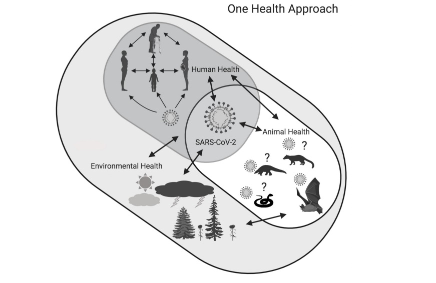
Figure 1. Set representation for the One Health approach in the context of coronavirus disease
2019 (COVID-19) caused by the SARS-CoV-2. Redrawn after Bonilla-Aldana et al6. Created with
BioRender.
Referencias bibliográficas
1. D.Gabe, C.Rodríguez, Vigliano, J.SanMartino, N.Wisner, et al. Mixomas cardiacos: correlacion anotomoclinica. Diponible en: https://www.sciencedirect.com/science/article/abs/pii/S0300893202766438
2. D.Muñoz, S.García, J.Páez, E.Hernández. Mixoma gigante de auricula derecha. Disponible en: https://www.sciencedirect.com/science/article/pii/S1134009613000168
3. T Zamora Bastidas, DE Maya Ruiz, M Rangel, N López Garzón. Mixoma: manifestaciones neurológicas y reumatológicas. Informe de casos. Revista Uruguaya de Cardiología. 2013; 28(1) 116-121. [ Links ]
4.N. López, M.Bermúdez Joaquí. Mixoma: manifestaciones neurológicas y reumatológicas. Informe de casos Tromboembolismo pulmonar secundario a mixoma gigante de aurícula derecha. Disponible en: https://www.sciencedirect.com/science/article/abs/pii/S0300893202766438
5. D.Calejero, M. González, R. Ortas, I. Ferreira. Trombolismo pulmonar secundario a mixoma gigante de auricula derecha. Disponible en: http://www.scielo.edu.uy/scielo.php?pid=S1688-04202013000100018&script=sci_arttext
6. I. Masuda, A.M. Ferreño, J. Pasca, G. Pereiro, H. Lastiri. Tumores cardíacos primarios. Mixoma auricular. Rev Fed Arg Cardiol, 33 (2004), pp. 196-204
7. F. Moreno Martínez, Á. Lagomasino Hidalgo, O. González Alfonso, I. Puig Reyes, R. Mirabal Rodríguez, O. López Bernal, et al. Mixoma auricular izquierdo pediculado con aspecto macroscópico de trombo calcificado. Rev Arg Cir Cardiovasc, 4 (2004/2005), pp. 251-255
8. R.P. Becker, S.P. Frangini, G.P. Arnaiz. Mixoma auricular izquierdo recurrente en niño de 2 años. Caso clínico. Rev Med Chile, 134 (2006), pp. 635-640
9. M. De Paula Vale, A. Freire Sobrinho, M. Vinícius Sales, M. Meirelles Teixeira, K. Chaves Cabral. Mixoma gigante em átrio esquerdo — Relato de caso. Rev Bras Cir Cardiovasc, 23 (2008), pp. 276-278
10. J.G. Lobo Filho, D.L. de Sá Sales, A.E.P. Pereira Borges, M.C. Leitão. Mixoma de átrio direito com prolapso para o ventrículo direito. Braz J Cardiovasc Surg, 21 (2006), pp. 217-220
11. F. Raila, P. Patel, B. Avera, J. Sigler. Echocardiographic diagnosis of left atrial myxoma. South Med J, 75 (1982), pp. 1120-1122
12. H. Hong, L. Yi, G. Shigong, Y. Xuezhong. Right atrial myxoma-induced syncope. Postgrad Med J, 87 (2011), pp. 438-439
13. O.B. Dike, S.S. Ajiduku, O.O. Omonua, L.L. Abdulkareem, W. Parsonage. A probable right atrial myxoma prolapsing through the tricuspid valve into the right ventricle: a case report. Cases Journal, 1 (2008), p. 386 Disponible en: http://www.casesjournal.com/content/1/1/38614
14. Enzo L. González, M.N. Pizzi, M.G. Caponi, C. Vigliano, M.D.P. Varela Otero, Dulbecco E., et al. Mixomas cardíacos: presentación clínica, resultados quirúrgicos y pronóstico a largo plazo. Rev Arg Cardiol, 78 (2010), pp. 108-113.

