English (pdf)
 Article in xml format
 Article references
 Send this article by e-mail
 Cited by SciELO
  Similars in
SciELO
Similars in
SciELO  uBio
uBio 
 Permalink
INTRODUCTION
Pomegranate (Punica granatum), Lythraceae, is an ancient deciduous shrub originative from Asia, expanded worldwide due to its fruits highly appreciated for its nutritious value 1), (2). The juice of pomegranate has been the subject of more than 250 studies where its benefical activity for cardivascular disease, cancer and arthritis is described (2). The root-bark, stem-bark, leaves and fruit peel of pomegranate contain tannins, flavonoids, lignans, triterpenoids and phytosterols, fatty acids, organic acids, phenolic acids 1),(3. Alkaloids found in pomegranate are pelletierine, pseudopelletierine, andN-methylpelletierine, sedridine, 2-(2′-hydroxypropyl)-∆1piperideine, 2-(2′-propenyl)-∆1piperideine, norpseudopelletierine, hygrine and norhygrine (root-bark), 3),(4.N-(2′,5′-dihydroxyphenyl)pyridinium chloride (leaves) 5, and pyrrolidine-type alkaloid punigratane (2,5-diheptyl-N-methylpyrrolidine) (fruit peel) 6. Besides alkaloids, indolamines, tryptamine, melatonin, and serotonin, were present in the extract of pomegranate fruit 7. The alkaloid pseudopelletierine (1) was chosen as example by authors 1 in order to show the importance of natural products and its impact in human health. Being a natural substance an organic compound, a chemical historical approach of the development of it is highly illustrative for graduate students, just as the fascinating book “Natural Products, Isolation, Structure Elucidation, History” by D. Sicker et al. exposes it 1. History of pseudopelletierine is described by authors in the book that included its isolation, purification, and structural elucidation, the last by means of historically developed chemical degradations and its historical synthetic approach. Finally, a set of modern spectral material, IR, UV MS and of course today’s current methods of NMR techniques spectra are available. Moreover, the book provides a supporting information book that explains chemical reactions for degradation and full synthesis of pseudopelletierine, as well as its biosynthesis. Also, the review of other alkaloids from pomegranate such as pelletierine, isopelletierine, methyl(iso)pelletierine, norpseudopelletierine, sedridine and hygrine are boarded by the authors of the book 1.
In this article, we just want to give a short collateral approach on the 13C NMR spectrum of pseudopelletierine 1, that could be illustrative for the reader.
THE 13C NMR SPECTRUM OF PSEUDOPELLETIERINE [1]
Analysis of chemical shifts based on tabled data
Figure 1 shows the APT 13C NMR spectrum 8 of compound 11. The spectrum shows six peaks, these are summarized in Table 1.
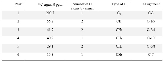
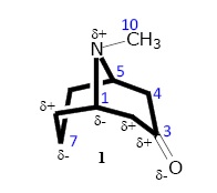
Peak 1 is according to the APT 13C NMR spectrum (Fig. 1) a quaternary carbon, because of its short height. In such plotting 1, this peak in anti-phase (downwards) and in this sense, it is regrouped with methylene peaks, being in phase (upwards) thus, peaks representing methyl and methine carbons.
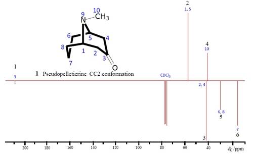
Fig. 1 Mimetic APT 13C NMR spectrum, pseudopelletierine (1) (constructed here in Chemwin® by authors, using the original spectrum from reference [1])
The difference between a DEPT 135 and an APT spectrum, is that in the first one, quaternaries disappear in comparison with the full normal 13C spectrum, and in the second one we can read all type of carbons independently of the number of hydrogens attached to it (0-3) . In this aspect, the APT spectrum is analogue to the JMOD 13C spectrum.
By inspecting 13C tabulated data 9, peak 1 falls in the range of carbonyl compounds (220 to 160 ppm, Table B10 9). The precise value of peak 1, d 209.7, permits to reduce the type of carbonyl to one pertaining to a ketone function, discarding thus all other types like esters, acids or aldehydes. Moreover, specific ketone spectral data can be referenced from table C190 9 where cyclohexanone (2) has a d 209.7, exactly the same value of C-3 in 1. This exact coincidence strongly suggests not only the ketone assignment for peak 1, but the cyclic feature of it too (a six-membered ring). Inspecting table C197 [9] we found compound 3, that is 2-oxopiperidine (2-piperidone, valerolactame). The latter possesses its carbonyl resonating at d 171.9 9, more shielded than C-3 in 1. Obviously, in 3, being nitrogen one-bond-distant neighbor of C=O, its Lewis’ basic character provides electronic shielding to carbonyl, consequently being its d shifted toward upfield. The fact that d of C=O in both 1 and 2 is that similar, makes us to think that the assignment of C=O in position 4 of piperidine in 1 (4-oxopiperidine) provokes no shielding at all over this carbon (because of its farness). Thus far, on the basis of tabled data, peak 1 permitted the proposal of 4-oxopiperidine portion (4-piperidone, 4), as possible structural fragment of 1.
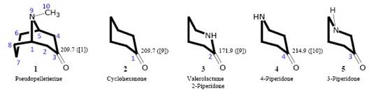
Compound 4 has a reported d of 214.9 ppm 10, giving an acceptable Dd(C=O, 4, 1) = 5.2 ppm with respect to the same carbon in 1. We haven´t found the reference for 13C data of the other structural possibility, 3-piperidone (5), in order to make a comparison with data of 1. However, we found in Table C177 ([9]), the values of chemical shift values for piperidine where the shielding effect of the heteroatom on C-2 is much higher than that on C-3 and C-4, being these two latter comparable (Dd = 1.9 ppm). This means that C-3 in 3-piperidone is close in d value to C-4 in 4-piperidone. As conclusion we deduce that 5 is also an acceptable structural proposal (besides 4) after analysis of peak 1 (Fig. 1).
Peak 2 in the APT 13C spectrum (Fig. 1) belongs to the phased signals (upwards) regrouping methyl and methine groups. At this stage of the analysis, we signal the symmetric character of the molecule, meaning by this a symmetry plan containing in itself C-7, N-9, and C-3 of 1, bisecting the molecule in two identical chained carbon atom series: C3-C2-C1-C8-C7 and C3-C4-C5-C6-C7 (the first one is the the specular image of the second one with chiral centers at 1S and 5R) where the couples 2 and 4, 1 and 5, and 6 and 8, possess identical NMR values since each carbon in the couple resonates at the same frequency. The spectral shape calls us for our attention since we can observe six different intensities (or heights) for the six signals, being the shortest one peak 1 and the tallest one peak 2. Table 1 shows peak 2 at d 55.8. Its high intensity shows this is a methyl group or a methine signal for two chemically equivalent (symmetrically disposed) methine carbon atoms. Table B5 [9] shows the range 60-40 ppm as including methylene, methyl and methine groups as saturated hydrocarbon portions. The methylene groups remain excluded since this is a phased (upwards) signal. The only two types of methyl groups included in this range are methoxy and methylamine groups (Table B5 [9]). From the bibliographic source, 1 has the molecular formula C9H15NO, derived from historical approach and from EIMS analysis with a molecular weight reflected by the molecular peak at m/z 153 1. Given the fact that the only oxygen of the formula corresponds to the 3-C=O group, then only the methylamine group should be considered between the range. Table C177 9 shows N-methylpiperidine with its methyl carbon resonating at d 47.7, value much less than peak 2 at 55.8 ppm. Based on the latter information, we can easily discard a methyl amine group, the only possible assignment for peak 2 being a methine (CH) group. Returning to table B5 9, we find a methine group trisubstituted by 3 carbons falling into the 60-40 ppm range. Also, in the same range can be found methine of alkynes, however, as can be stated from the molecular formula, and employing the index of hydrogen deficiency 11 (this index is the sum of the number of rings, the number of double bonds, and twice the number of triple bonds 11), the calculated index for 1 is 3.
Index = #C - #H/2 - #X/2 + #N/2 + 1
Index = 9 - 15/2 - 0/2 + ½ + 1
Index = 9 - 7 +1 = 3
Interpreting the index 3 for 1, we have one double bond in 3-C=O, and since there are no alkene carbons (140-80 ppm, see Fig. 1 and Table 1), there are not C=C. The 2 remaining units could well correspond to C C. However, this is contrary to the suggested 4- or 3-pipiridone, already established from analysis of peak 1. Consequently, the second of 3 units of the index corresponds to such six-membered cycle. The last unit of the 3 of the index cannot be other than a second ring, given the fact of the absence of more double bonds and that we have 9 carbons, and 5 belong to piperidone. As conclusion, 1 is a bicyclic compound. Continuing in the range 60-40 ppm in Table B5 9, we find (C)2CHCOX; X:C, O, N, this means that the methine is directly linked to two carbons and to a carbonyl. This latter can be ketone, ester or amide. In 1 we have a cyclic ketone (4- or 3-pipiridone), hence, ester and amide are discarded. 6 and 7 show the two possible piperidone portions of 1 when methines are directly linked to carbonyl.
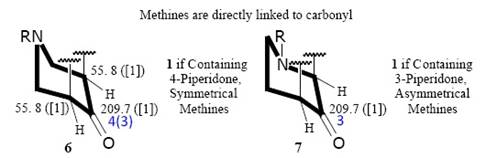
According to the graphic above (7), the option 3-piperidone as the first cycle of 1 is discarded cause methines are not symmetrical. On the contrary, due to the symmetrical disposition of methines at 55.8 pm, 4-piperidone (6) is acceptable as the first cycle of 1.
Finally in table B5 [9] we find (C)2CH-N in the range 60-40 ppm, it means a methine linked to two carbons and one nitrogen. Since we have established peak 2 as methine and that its high intensity suggests two chemically equivalent carbons (same d, symmetrically disposed), we are assigning to peak 2 two carbons, mirror images from each other, each in one of two identical moieties of the molecule. Having been discarded 7 (3-piperidone in 1), 8 shows the remaining 4-piperidone when methines are directly linked to N but not to carbonyl. Of course, these latter are symmetrically disposed.
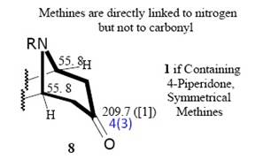
Since in this discussion we placed firstly methines in a of carbonyl (HC-C=O, 6, 6’), then the symmetrical methylene groups are placed in b of carbonyl of 4-piperidone. If we place methines in b of carbonyl, then methylene groups are in a of carbonyl (H 2 C-C=O, 8, 8’). Summarizing, depending of the position of the fourth carbon from the 4 remaining after assignment of the first five to 4-piperidone, we have four possible structures for 1, 6, 6’, 8 and 8’. We can at this point reassure the apparition of the second ring of bicyclic 1.
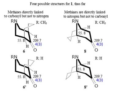
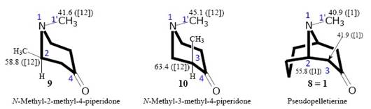
Peak 4. Peak 3 and peak4 in the APT 13C spectrum (Fig.1) are downwards, and upwards, meaning thus a methylene and a methyl group, respectively. The alternative of a methine for peak 4 is discarded from the summing of the number of protons, thus far, it means that in any of the 4 possible structures for 1, 6, 6’, 8 and 8’, we count 15 protons, in accordance with the molecular formula C9H15NO. 6 and 8 show the N-methyl-4-piperidone nucleus (containing the N-CH3 group). It’s unconceivable to propose a N-CH group. On the other hand, 6’ and 8’ show the 4-piperidone nucleus (containing the N-H group). Hence, the counting of protons in 6 and 8 is as follows: 5CH2 + 2CH + 1CH3N= C8H15N. The counting of protons in 6’ and 8’ is 6CH2 + 2CH + NH = C8H15N. In all four alternative structures for 1 (6, 6’, 8 and 8’) the maximum number of methines (CH) is 2 (already assigned to peak 2), consequently, peak 4 is not a methine but a methyl group pertaining to the 9 carbon atoms in the molecular formula, where 5 belong to the nucleus 4-piperidone of 1. The chemical shift value of N-CH3 in 1 is d 40.9 (Table 1) that compared with the d 45.9 of methyl in N-methyl-4-piperidine (Table C177 9) presents a small difference of 5 ppm, interpreted by us as a proven assignment of the group N-CH3 in 1. Moreover, we found a bibliographic source (12 that gives 13C data for N-methyl-4-piperidones. In Table I 12, the N-CH3 carbon receives the value d 41.6 (C-1’), in structure V (N-methyl-2-methyl-4-piperidone, see compound 9 here), very close to d 40.9 in 1 (Dd = 0.7 ppm). In that very same compound (V, 9) carbon 2 (in alpha of amine) shows a d 58.8 comparable to 55.8 ppm of C-2 in 1 (Dd = 3.0 ppm). On the other hand, the same source shows structure VIa or N-methyl-3-methyl-4-piperidone, see compound 10 here), for which C-3 has a value d 63.4, much different from d 55.8 in 1 (Dd = 7.6 ppm), and C-1’ with d 45.1 much different from C-1’ in 1 (40.9, Dd = 4.2 ppm), and finally very different from C-3 in 1 (41.9, Dd = 21.5 ppm). In conclusion, these data from source 12 permitted us to define the position of the methines of junction of rings, of the portion N-methyl-4-piperidone of 1 as C-2 (or positioned in a of amine, and not on a of carbonyl), this under the optics that substituent in C-2 is an alkyl group, and that the CH3 group in 9 is magnetically comparable to the group -CH2-CH2- in 1. Since we have defined the location of the only methyl group as N-CH3 and that the two methines, rings’ junction carbons (C-2 of N-methyl-4-piperidone of 1) are placed in a of amine and not in a of carbonyl, then, we can discard structures 6, 6’ and 8’. This puts structure 8 as the final structure of 1.
Peaks 3, 5 and 6. Until here we have found the structure of pseudopelletierine, by using solely the APT 13C NMR spectrum as a spectroscopic tool (assisted by mass data). Demonstrating that for the present example, all other resources of NMR to solve structures can be saved, at least as an initial approach. However, to achieve the completion of the full assignment of signals of 1, the study of the chemical shift values of the 3 methylene groups in ring B must be undertaken, as well as the description of two methylene groups in ring A.
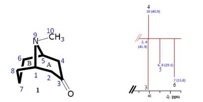
From 8, we know that the structural complement of the N-methyl-piperidone moiety, or ring A of 1 is the fused ring B composed by the bridge atoms 1, 5 and 9, and complemented by atoms 6, 7 and 8. Here, C-6 and C-8, and C.-1 and C-5 are symmetrical (mirror images). The symmetrical methines C-1 and C-5 were already worked. 9 is nitrogen. Then in ring B, symmetrical C-6 and C-8 have one same 13C signal (a first of three CH2 groups in Fig. 1). The second signal methylene corresponds to C-7. The third peak downwards must correspond to two equivalent methylene groups not described yet, namely C-2 and C-4 in ring A. A priori, it is easy to assign the three peaks, 3, 5 and 6, on the basis of the d value of each. It is obvious that the most shielded nucleus is C-7 (peak 6, 15.8 ppm), this due to the extreme farness from highly electronegative atoms, it is worth to say, N and C=O. Somehow more de-shielded is peak 5 (29.1 ppm), this can be assigned the couple C-6/C-8. This because of its closeness to nitrogen, being placed in b of N. By default, and due to its placement between two strong de-shielding factors, the carbonyl and the amine groups, the couple C-2/C-4, is assigned to peak 3 (41.9 ppm). This couple is placed in a of C=O and in b of N. Nevertheless, to be orthodox, we need to check this premature conclusions by examining tabled data. Peak 3 (41.9 ppm) is according to Table B5 [9] (60-40 ppm range) a methylene between 2 carbons (a corroborating fact, since C-2/C-4 are between C-3 and C-1/C-5). Secondly, Table B5 shows a methylene directly bonded to nitrogen, (let’s say in a of N). Thirdly, these symmetrical methylene groups can be placed in a of C=O (Table B5 [9]). There is no contradiction in these three assignments grosso modo. Moreover, Table C190 [9], shows methylene in a of C=O of cyclohexanone with d 41.5 ppm (Dd = 0.4 ppm). To be more precise, reference 12 shows compound IV, N-methyl-piperidone, with d 40.9 for the couple of methylenes C-3/C-5. Peak 5 (29.1 ppm), this d value falls into the range 40-20 ppm in Table B5 [9] that firstly shows a methylene directly bonded to two other carbons, it is just the case for C-6/C-8 in ring B of 1 (between methines 1/5 and methylene C-7), there are no more data in this table (B5 [9]). Table C177 [9] shows C-3 at d 26.4 in N-methylpiperidine (Dd = 2.7 ppm) corroborating the a priori assignment. Peak 6 (15.8 ppm) falls in the range 20-0 ppm in Table B5 [9], corresponding to aliphatic methylene (between C-6 and C-8). Table C177 [9] shows C-4 at d 26.4 in N-methylpiperidine, this value is not close to 15.8 ppm of C-7 in 1. We found a reference 13, where different measurements on cyclooctanone-1,2-D 2 were performed, giving values like 25.18, 24.88, 24.78 and 24.76 ppm for C-5 of cyclooctanone. These two values, C-4 (N-methylpiperidine) and C-5 (cyclooctanone) are more alike between them than with C-7 (15.8 ppm) in 1. Thus, in order to propose an explanation for the value 15.8 ppm of the highly shielded C-7 of 1, we deduce that the combination of N-methylpiperidine and cyclooctanone made possible such shielding on C-7 of 1. The following graphic proposing partial electronic densities generated by carbonyl as a kind of driving force could justify an electronic accumulation over the C-7 nucleus of 1. In this graphic the driving force exerted by carbonyl would be superior to that exerted by nitrogen, in spite of the through-the-bonds distance. The value of d 20.8 (C-7) in 1, was reported in 1 as a 13C-NMR signal predicted by ChemBio-Draw.