


Servicios Personalizados
Revista

Articulo
 Inglés (pdf)
 Articulo en XML
 Referencias del artículo
 Enviar articulo por email
Indicadores
 Citado por SciELO
 Links relacionados
 Similares en
SciELO
Similares en
SciELO  uBio
uBio Compartir

 Permalink
Revista Boliviana de Química
versión On-line ISSN 0250-5460
Rev. Bol. Quim v.29 n.1 La Paz 2012
ARTÍCULO ORIGINAL
APPLICATION OF FERRIPROTOPORPHYRIN BIOCRYSTALLIZATION INHIBITION TEST TO FIND ANTIPLASMODIAL COMPOUNDS IN THE FLORA OF THE "ZONGO VALLEY" - BOLIVIA
Sandra L. Ibañez Calero12*, Valérie Jullian3, Séverine Maurel3, Lia R. Chavez de Michel4 Jose A. Bravo16, Alberto Gimenez5, Michel Sauvain3
1 Instituto de Investigaciones Químicas, Universidad Mayor de San Andrés (UMSA), Casilla 303, La Paz, Bolivia. 2220 Shangri La Ln, Pittsburgh, PA, 15239. 3 UMR 152, Institut de Recherche pour le Développement - Université Paul Sabatier, Faculté des Sciences Pharmaceutiques 31062 Toulouse cedex 09, France. 4 Herbario Nacional de Bolivia (UMSA), Casilla 10077, La Paz, Bolivia. 5 Instituto de Investigaciones Fármaco-Bioquímica (UMSA), Casilla 3239, La Paz, Bolivia.6 Instituto de Investigaciones en Productos Naturales (IIPN), Carrera de Ciencias Químicas, Universidad Mayor de San Andrés, Calle Andrés Bello y Calle 27 Cota Cota, Edificio FCPN, 2° piso, La Paz- Bolivia.
Keywords: Ferriprotoporphyrin biocrystallization inhibition test, antiplasmodial, Brachyotum microdon, Bolivia
ABSTRACT
Among the different tests or chemical reactions that explain the possible inhibition mechanisms of Plasmodium falciparum growth, we have chosen the Ferriprotoporphyrin biocrystallization inhibition test (FBIT) to evaluate the pharmacological properties of 65 plants from the Bolivian flora. A bioguided phytochemical separation of Brachyotum microdon, one of the most active plant, was sought using the FBIT test. The isolated active compounds belong to two types of metabolites the fatty alcohols and the triterpenes. Among the triterpenes, we have p-sitosterol (1), oleanolic acid (2), ursolic acid (3) and corosolic acid (4) as sources of the activity on FBIT.
INTRODUCTION
Malaria is one of the most troublesome tropical diseases that kills more people than any other communicable disease except tuberculosis and AIDS. In 2009, 225 million malaria cases have been reported and nearly one million deaths. [1]. The remerging of this widespread parasitic disease in areas where it was previously under control or eradicated and the appearance of drug-resistant malaria parasites have aroused in the search for new leads for antimalarial drugs [2, 3, 4, 5]. The latest developments of molecular biology in the parasite [6, 7, 8] have provided new biological drug targets and modes of action [5, 9, 10]. Malaria parasites digest hemoglobin during their intra erythrocytic life cycle to obtain the required amino acids [11]. The by-product of this digestion is the release of the toxic hemin (ferriprotoporphyrin IX), which is detoxified by the parasite via formation of an insoluble crystal (hemozoin) [12, 13]. There are indications that this malarial crystal is identical to synthetic p-hematin [14, 15]. In the Ferriprotoporphyrin Biocristallization Inhibition Test (FBIT), the aim is to reproduce in vitro the biocrystallization reaction and to test the ability of synthetic drugs [16, 17] or natural extracts [18, 19, 20] to inhibit this detoxification pathway.
Biological chemodiversity continues to play an important role in the search for leads for antimalarial drugs since the majority of the existing antimalarial chemotherapeutic agents are based on natural products. Active compounds, with a variety of structures, have been isolated from plants, bacteria, fungi, marine and fresh water organisms [21, 22, 23, 24].
Bolivia, located at the center of South America, has different ecosystems each of them with a specific climate, altitude and soil. One of these ecosystems is the Zongo Valley, which is a high, moist, tropical location in the south west of the department of La Paz - Bolivia [25, 26]. The valley presents around 109 families and 518 species [27]. We assayed part of this biodiversity using the FBIT and the active species were submitted to the Plasmodium falciparum test.
EXPERIMENTAL
2.1. General
Optical rotations were measured in a Perkin- Elmer 241 digital polarimeter at 25°C and at 589 nm. IR spectra were recorded on a Perkin- Elmer Spectrum One FTIR. NMR spectra were recorded on Brucker, AMX 400 and Avance 500. DCI and EI were performed using a Nermag R10- 10 mass spectrometer. HPLC was performed using a Waters Xterra RP18 column of 10 |im, 19 x 250 mm. All supports and reagents used in this work were obtained from Merck and Sigma.
2.2. Plant Species
Plant species were collected in the "Valle de Zongo" in October 2001, February and May 2002. The collection started near the Zongo Dam at altitude 4715 m a.s.l. (68°05'02 " longitude and 16°15'02 " latitude) and ended near the Huaji Hydroelectric Power Station at 941 m a.s.l. (67°55'04 " longitude and 16°00'05 " latitude). All species were identified and deposited in the Bolivian National Herbarium, La Paz.
2.3. Extracts
The air-dried specimens were separated into their different organs, grinded and extracted with ethanol (200 mg/ml) for 24 hr. The dried extracts were tested in FBIT and only the active species were submitted to the P. falciparum test.
2.4. FBIT
The ability of the extracts and fractions to inhibit ferriprotoporphyrin IX (FPIX) biocrystallization was assessed by following the protocols previously reported by our group [27, 28, 29]. FBIT is useful for the detection of compounds, presented in plant extracts, which interfere with FPIX biomineralization. It has been observed in Deharo et al. [29], that colored components do not interfere with the measurements of absorbances. In Ibanez-Calero et al. [28], we used FBIT to perform a bioguided isolation of a new anthraquinone from Rumex obtusifolius.
2.5. Plasmodium falciparum Test
Cultures of F32-Tanzania (chloroquine sensitive) strains of P. falciparum were maintained according to the method of Trager and Jensen [30].
2.6. Extraction and active compounds isolation on Brachyotum microdon Brachyotum microdon
The plant studied belongs to the Myrtales order, to the Melastomataceae family, to the Brachyotum genus and to the Brachyotum microdon specie. Its binomial name is Brachyotum microdon (Naudin) Triana. It is commonly known as the Rhododendrons of the Andes because of its splendor when in flower. Brachyotum microdon is a large shrub, erect and loaded with fuchsia- like flowers. Their leaves are simple, commonly opposite and decussate with one of a pair slightly smaller than other. They are rarely verticillate or alternate by abortion of one of a pair. They usually present 1 to 5 secondary veins on each side of the midvein and numerous tertiary veins, parallel, and connecting secondary veins and midvein. The flowers of Brachyotum microdon are pendant, bisexual, actinomorphic, with 4 to 5 merous and with opposite bracteoles usually caducous. They are tubular (10 to 15mm long) and present a red hypanthium. The calyx is valvate with 3-5 lobes. The petals, 3 to 6 as the number of sepals, are distinct and imbricate. Brachyotum microdon has stamina nectaries on the dorsal surface of the anther connective. Nectary stomata are distributed in lines along the proximal third of the connective and are placed above large intercellular spaces of the parenchyma. The vascular bundle in the connective is large, paralleling the epidermis by sometimes only three layers of parenchyma cells. The anthers are typically two, celled, with 1 or 2 apical pores. The flowers have one pistil, one style and a minute stigma. The ovary is commonly inferior with 3-5 locules with various ovules. The fruits are dry or fleshy capsuled with small seeds [31, 32]. Figure #1 presents a picture of the collected plant.
Extraction and active compounds isolation
Dried leaves (2 kg) of Brachyotum microdon were successively extracted by 48 hr maceration at room temperature, 3 times with petroleum ether (6 L), dichloro methane (7 L) and MeOH (7 L). The dichloro methane extract concentrated the activity in FBIT; it had 64 % inhibition at 1.25 mg/ml. The active fraction (31 g) was fractionated on a vacuum liquid chromatography column (VLC) eluted with petroleum ether - dichloromethane - EtOAc- MeOH mixtures of increasing polarity to yield 13 fractions.
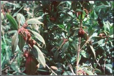
Figure 1. Brachyotum microdon, Melastomataceae
Fractions 7 to 9 (IC50< 0.25 mg/ml, 3487 mg) were joined and chromatographed on a Flash column eluted with CH2Cl2 - AcOEt - MeOH mixtures to yield 11 fractions. Fraction 3 provided a mixture of fatty alcohols (18.6 mg, IC50: 0.2 mg/ml) after a PTLC with CHQ3 (Rf= 0.16 an orange spot visualized with H2SO4, 25%). This mixture corresponds to C34H70O, C32H66O, C30H62O, C28H58O and C26H54O. Fraction 7 was chromatographed on silica gel with CH2Cl2- AcOEt (95:5) to give 2 sub-fractions. From sub-fraction 2 and after crystallization from methanol, p-sitosterol (1) (15mg, IC50: 0.2 mg/ml) was obtained. Fraction 9 (82.2 mg, IC50: 0.19 mg/ml) was subjected to a size exclusion chromatography with sephadex LH-20 to give 5 sub-fractions. Sub-fraction 3 (34 mg) was further purified by PTLC (CH2«2, Rf= 0.18 a red spot visualized with H2SO4, 25%) to afford oleanolic acid (2) (9 mg, IC50: 0.61 mg/ml).
From the VLC, fraction 10 was further fractionated on a Flash column eluted with c-hexane - AcOEt mixtures to give 7 sub-fractions. From sub-fraction 2, ursolic acid (3) (105.3 mg, IC50: 1.6 mg/ml) was obtained after crystallization from chloroform. Sub-fraction 5 (133.1 mg) was purified with a silica gel Flash column providing more ursolic acid (30mg) eluted with c-hexane - AcOEt (80:20) and a mixture of 2 diasterotopic triterpenes (9 mg), which were separated by HPLC eluted with MeOH: H2O-TFA 0.1% (68: 32). The main triterpene (4) had a retention time of 37.7' (4 mg, IC50: 0.2 mg/ml). The structures of the known compounds were correlated with literature [33, 34, 35, 36].
Corosolic acid(4). White powder, [a]D25 + 25° (c 0.10, CH3OH); IR (KBr) vmax 3414, 2928, 2859, 1690, 1457, 1384, 961, 802, 742, 617; 1H NMR (CDCl3-CD3OD [4:1]): 8 5.20 (1H, t, 3.6 Hz, H-12), 3.63 (1H, m, H-2), 2.91 (1H, d, J= 9.5 Hz, H-3), 2.18 (1H, d, 10.5 Hz, H-18), 1.98 (1H, ddd, J= 12.5, 12.5, 5 Hz, H-16), 1.97 (1H, d, J=
12.5 Hz, H-1'), 1.93 (2H, dd, J= 8.5, 4.0 Hz, H-11), 1.86 (1H, ddd, J= 12.5, 12.5, 5 Hz, H-15), 1.68 (1H, dd, J= 14.0, 14.0 Hz, H-22), 1.63 (1H, dd, J= 12.5, 5 Hz, H-16'), 1.63 (1H, dd, J= 14.0, 4.5 Hz, H-22'), 1.56 (1H, d, 8.5 Hz, H-9), 1.54 (1H, dd, J= 9.0, 8.5 Hz, H-7), 1.52 (1H, d, J= 13.8 Hz, H-21'), 1.50 (1H, dddd, J= 10.0,10.0,10.0, 5.0 Hz, H-6), 1.38 (1H, dd J= 12.5, 12.5 Hz, H-21), 1.35 (1H, m, H-6'), 1.35 (1H, m, H-7'), 1.34 (1H, m, H-19), 1.08 (3H, s, H-27), 1.06 (1H, m, H-15'), 0.98 (3H, s, H-23), 0.98 (3H, s, H-25), 0.98 (1H, m, H-20), 0.93 (3H, d, J= 6.5 Hz, H-30), 0.90 (1H, dd, J= 12.5, 10.5 Hz, H-1), 0.85 (3H, d, J= 6.5 Hz, H-29), 0.79 (6H, s, H-24, H-26), 0.78 (1H, d, J= 10.0 Hz, H-5); 13C NMR (CDCl3-CD3OD [4:1]): 8 180.5 (C, C-28), 138.7 (C, C-13), 125.2 (CH, C-12), 83.4 (CH, C-3), 68.5 (CH, C-2), 55.3 (CH, C-5), 52.8 (CH, C-18), 47.8 (C, C-17), 47.5 (CH, C-9), 46.5 (CH2, C-1), 42.1 (C, C-14), 39.5 (C, C-8), 39.2 (C, C-4), 39.1 (CH, C-19), 38.9 (CH, C-20), 38.0 (C, C-10), 36.8 (CH2, C-22), 32.9 (CH2, C-7), 30.6 (CH2, C-21), 28.5 (CH3, C-23), 28.0 (CH2, C-15), 24.2 (CH2, C-16), 23.4 (CH3, C-27), 23.3 (CH2, C-11), 21.0 (CH3, C-30), 18.3 (CH2, C-6), 16.8 (CH3, C-29), 16.8 (CH3, C-24),
16.6 (CH3, C-26), 16.5 (CH3, C-25); DCIMS, m/z 490 [M+NH4]+ (100), 473 [M+H]+ (9), 455 (5), 446 (31), 428 (3), 248 (3); EIMS, m/z 248 (100), 233 (1), 203 (22), 189 (9).
2.7. Structure-Activity proposal
Portela et al. have demonstrated that quinolic and xanthonic type molecules interact with hemin chloride and stabilize it thanks to large range interactions determined by their complementary electrostatic profiles [37]. Following Portella's work, we have published a structure-activity proposal for a new antraquinone isolated from Rumex obtusifolius and active in the FBIT test [28]. In these works, the electronic centers of the active compound will interact with the opposite poles found in the hematin (hemin chloride crystal). In the hemin chloride, the most negative potential is found in the position occupied by the iron in the tetrapyrrolic system while the most positive site is found at the propionic groups. One of the necessary characteristics that active compounds should have is the presence of a null or positive potential all along the molecule or the presence of an aromatic cycle. In this publication, we present a structure-activity relationship proposal to understand the fashion in which the acidic triterpenes, isolated in this work, would interact with hemin chloride.
RESULTS AND DISCUSSION
3.1. Extracts
We tested on FBIT 155 extracts obtained from 65 collected plants. Among these plants, it is important to emphasize that the leaves of Brachyotum microdon (Melastomataceae) showed an important inhibition in the FBIT (63 % inh. at 2.5 mg/ml) but no activity towards P. falciparum. However, the flowers of this plant presented the highest activity against P. falciparum in vitro (97 % inh. at 10 |g/ml). The very low amount of dried crude extract of Brachyotum's flowers makes the isolation of active compounds difficult. We decided to start the bioguided separation of Brachyotum microdon since to our knowledge it had never been previously studied.
3.2. Active compounds from Brachyotum microdon
Following a bioguided isolation process using FBIT, we obtained a mixture of fatty alcohols, p-sitosterol (1), oleanolic acid (2), ursolic acid (3) and corosolic acid (4). The structures of the known compound were correlated with literature [33, 34, 35, 36].
The biological results of the isolated compounds against FBIT and Plasmodium falciparum are shown in Table 2.
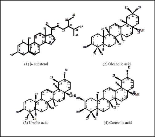
Table 2. Biological results of the isolated compounds against FBIT and Plasmodium falciparum
| COMPOUND | FBIT IC50 (mg/ml) | P. falciparum IC50 (µg/ml) |
| Fatty alcohols | 0.2 | NT1 |
| p-sitosterol | 0.2 | NT1 |
| Oleanolic acid | 0.6 | 96 |
| Ursolic acid | 1.6 | 59 |
| Corosolic acid | 0.2 | 100 |
| Quinine2 | 0.03 | NT1 |
| Chloroquine2 | 0.016 | 0.006 |
'NT: not tested 2:control compounds
As shown in Table 2, corosolic acid, p-sitosterol and the mixture of fatty alcohols gave the most important results in the FBIT, 0.2 mg/ml. In addition, corosolic acid gave an IC50= 100 |g/ml against P. falciparum in vitro. There are no previous reports on corosolic acid's FBIT activity or on its low antiplasmodial property. On the other hand, the antimalarial activities of fatty alcohols against different types of Plasmodium species were previously reported [38,39, 40]. The known ursolic and oleanolic acid were reported to exhibit antiplasmodial activity [41, 42]. Oleanolic acid gave in our work an IC50= 96 |g/ml against P. falciparum in vitro which is in agreement with the reported low activity against both chloroquine - resistant and - sensitive P. falciparum with mean IC50 values of 88.8 |g/ml and 70.6 |g/ml, respectively [43]. In addition, for ursolic acid we obtained an IC50= 59 |g/ml against P. falciparum in vitro which is close to the reported values, IC50= 37 |g/ml (against K1) and IC50= 28 |g/ml (against T9-96) [43]. Furthermore, interesting results against P. berghei in mice were obtained by Amusan et al. for ursolic acid [44]. This triterpene produced a 97% suppression of parasitemia and a mean survival period of 25 days at 60 mg/Kg/day.
The acidic triterpenes presented in this work have been previously reported with a vast variety of biological properties [45, 46]. Ursolic acid has shown antileishmanial properties [47, 48, 49], antiprotozoal attributes [47, 41], antibacterial characteristics [50, 51, 52], antidiabetic activities [53, 54], antifungal property [55], anti-inflammatory qualities [56, 57], anti-HIV feature [58] and antioxidant activities [59, 60, 61]. There is also a large anticancer and anti-tumor research directed to different targeted organs [62, 63, 64, 65, 66, 67, 68, 69, 70, 71, 61, 72, 57]. Ursolic acid has also been presented as a cough suppressant [73], blood circulation promoter [74] and as a pancreatic lipase inhibitor [75].
Oleanolic acid has also been reported with almost the same attributes than ursolic acid. It has antileishmanial properties [48, 47], antibacterial characteristics [76, 50, 51, 52], antioxidant activities [77, 60], anti-inflamatory quality [78], antiprotozoal attribute [47], anti-HIV feature [58], antidiabetic property [53] and antiatherosclerotic characteristic [79]. There are also several anticancer and anti-tumor publications with this triterpene [80, 67, 71, 81, 82, 61, 72]. In addition, oleanolic acid can improve lungs function in pulmonary treatments [77] and improve glucose tolerance [83]. It is also a photoprotector [84], has antiobese properties [83] and could have a hypolipidemic effect [85].
Corosolic acid has also many biological attributes. There are reports about its antidiabetic activities [86, 54, 87, 88, 89, 90], anticancer/anti-tumor properties [91, 92, 93, 94, 95], antiobese characteristics (86, 46], anti-inflamatory properties [96, 97] and antioxidant feature [97]. It has also been presented that corosolic acid has anabolic effects [98], has effects on the cholesterol absorption in diabetes studies [99], enhances antibiotic's activity [100], inhibits pancreatic lipase [75] and could prevent atherosclerosis [97].
3.3. Structure-Activity proposal
In this work, we used the FBIT to find compounds that could react with hemin chloride; therefore, inhibiting its crystallization to p-hematin. We isolated a series of acidic triterpenes that are active in the FBIT. The mechanism of this inhibition is still unclear; based on recent publications we have presented a proposal of a triterpene- heme complex formation [37, 28]. The proposed mechanism of action has two steps; first the attraction and stabilization of the hemin by the triterpene through electrostatic interactions and second the formation of the complex through a chelating ligand between the active compound and the hemin. This proposal is close to the two steps mechanism presented by Mullie et al. [101]. The first step in Mullie's work, using ursolic acid derivatives as the active molecules, consists on the stacking of a hydrophobic structure to hemin. The second step is an additive protection of the hemin ferric iron by the ursolic acid's hydroxyphenil substituents through steric hindrance. This second step is dependent and enhanced by the derivative moiety added to the ursolic acid main skeleton. In addition, the importance of the hydrophilic framework attached to the triterpene and the possible interaction implicating binding of the active compounds to p- hematin is also reported by Gnoatto et al. [102].
It is possible to propose an electronic behavior in these triterpenes based on their partial electronic densities. At the extremes of the molecules, at the acid and alcohol groups, two zones of negative potentials are localized. In addition, in the center of the molecules, there is a positive potential that joins the null potential generated by the double bond. These electronic centers interact with the opposite electrostatic forces found in the synthetic hemin. The negative potential in the hemin chloride will be stabilized by the positive and null zones found in the triterpenes while the positive potential in the hemin will interact with the negative ones found in the triterpenes. Once this stabilizing interaction is reached, the carboxylic moiety of the triterpenes reacts with the iron in the synthetic hemin displacing the chlorure (Cl-) through an electronic attack. In this fashion the coordination bond FeOCO is formed. Figure #2 shows (A) the electronic stabilization of hemin chloride by a triterpene and (B) the complex formed by hemin chloride-triterpene. We present this proposal with corosolic acid since it was the most active triterpene in the FBIT test.
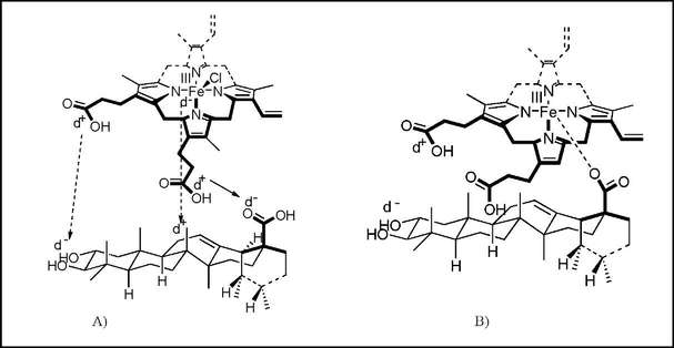
Figure 2. A) Stabilization of hemin chloride by corosolic acid. B) Complex of hemin- corosolic acid
The low antimalarial activities of the triterpenes presented in this work could be explained following Sonnet et al.'s work [103]. They studied the abilities of antimalarial chelating compounds and ursolic acid derivatives to inhibit p-hematin formation. They concluded that the iron chelation is not the main driving force behind the inhibition of the heme crystallization and heme degradation; therefore, supporting the need of hydrophilic substituents to enhance the molecule's existing activity. The structural differences among the presented triterpenes may cause different in vitro and in vivo antiplasmodial activities due to the possible following factors: changes on compound solubility (in culture media and in lipid membranes), altered target site interaction and different compound's electronic profiles.
There are no known previous reports of Brachyotum microdon, making oleanolic acid, ursolic acid and corosolic acid the firsts compounds isolated from this species. Neither, corosolic acid or oleanolic acid has been presented as inhibitors of the biocrystallization of FPIX. In addition, corosolic acid has not been previously reported to exhibit an activity against P. falciparum.
More recently, it was reported that piperazine derivatives of ursolic acid have important activities against Plasmodium falciparum CQ-resistant strain (IC50= 78- 167 nM) [102]. Furthermore, Suksamran et al. have also publish that the derivatives of oleanolic acid and ursolic acid with a p- coumarate moite at the C-27 position have enhanced antiplasmodial activities in vitro (IC50= 2.9 µg/ml for both acidic triterpenoids) [104]. In addition, a series of ursolic acid derivatives with a variety of substituents at C-3 and C-28 position were synthesized showing increased activity against P. falciparum [105]. These findings leave open the possibilities to prepare a series of derivatives of the three isolated acidic triterpenes, particularly of corosolic acid and oleanolic acid, for further FBIT and antiplasmodial studies.
ACKNOWLEDGEMENTS
National Science Grant from ACI Pal +, Research Ministry of France.
Notes
REFERENCES
1. World Health Organization. World malaria report 2010. Geneva, Switzerland. [ Links ]
2. C. W. Wright;Nat. Prod. Rep. 2010, 27, 961- 968. [ Links ]
3. K. C. Peach; R. G. Linington;Future Med. Chem. 2009, 1, 593- 617. [ Links ]
4. K. Papireddy; M. Smilkstein; J. X. Kelly;Shweta; S. M. Salem; M. Alhamadsheh; S. W. Haynes; G. L. Challis; K. A. Reynolds;Med. Chem. 2011, 54, 5296-5306. [ Links ]
5. A. Bell;Curr. Pharm. Des. 2011, Jul 5.(EPub ahead of print, PMID 21728986). [ Links ]
6. E. Hanssen; C. Knoechel; N. Klonis; N. Abu-Bakar; S. Deed; M. LeGros; C. Larabell; L. Tilley;J. Struct. Biol. 2011, 173, 161- 168. [ Links ]
7. B. Gupta; G. Awasthi; A. Das;Parasitol. Res. 2010, 107, 495- 499. [ Links ]
8. A. Bahl; B. Brunks; R. L. Coppel; J. Crabtree; S. J. Diskin; M. J. Fraunholz; G. R. Grant; D. Gupta; R. L. Huestis; J. C. Kissinger; P. Labo; L. Li; S. K. McWeeney; A. J. Milgram; D. S. Roos; J. Schug; C. J. Stoeckert Jr.; Nucleic Acids Res. 2002, 30, 87- 90.
9. C. Moneriz; J. Mestres; J. M. Bautista; A. Diez; A. Puyet;FEBS J.2011, 278, 2951-2961.
10. D. A. Fidock; P. J. Rosenthal; S. L. Croft; R. Brun; S. Nwaka; Nature reviews 2004, 3, 509- 520. [ Links ]
11. D. E. Goldberg; A. F. G. Slater; Parasitol. Today 1992, 8, 280- 283. [ Links ]
12. T. J. Egan; Molecular and Biochemical Parasitology 2008, 157, 127- 136.
13. J. Ziegler; R. Linck; D. W. Wright; Current Medicinal Chemistry 2001, 8, 171- 189.
14. T. J. Egan; Exp. Opin. Ther. patents 2001, 11, 185- 209.
15. S. Pagola; P. W. Stephens; D. S. Bohle; A. D. Kosar; S. K. Madsen; Nature 2000, 404, 307- 310.
16. S. Pérez-Silanes; L. Berrade; R. N. García-Sánchez; A. Mendoza; S. Galiano; B. M. Pérez-Solórzano; J. J. Nogal-Ruiz; A. R. Martínez-Fernández AR; I. Aldana; A. Monge; Molecules 2009, 14, 4120- 4135.
17. E. del Olmo; M. G. Armas; M. Ybarra; J. L. López; P. Oporto; A. Giménez; E. Deharo; A. San Feliciano;Bioorg. Med. Chem. Lett. 2003, 13, 2769- 2772.
18. G. Garavito; J. Rincón; L. Arteaga; Y. Hata; G. Bourdy; A. Gimenez; R. Pinzón; E. Deharo;J. Ethnopharmacol.2006, 107, 460- 462.
19. G. Bourdy; P. Oporto; A. Gimenez; E. Deharo;J. Ethnopharmacol. 2004, 93, 269- 277.
20. R. Baelmans; E. Deharo; G. Bourdy; V. Muñoz; C. Quenevo; M. Sauvain; H. Ginsburg; J. Ethnopharmacol. 2000, 73, 271- 275.
21. A. B. Oliveira; M. F. Dolabela; F. C. Braga; R. L. Jácome; F. P. Varotti; M. M. Póvoa;An. Acad. Bras. Cienc. 2009, 81, 715- 740.
22. R. Batista; J. Silva Ade Jr.; A. B. de Oliveira;Molecules 2009, 14, 3037- 3072.
23. K. Gademann; J. Kobylinska;Chem. Rec.2009, 9, 187- 198.
24. K. Kaur; M. Jain; T. Kaur; R. Jain;Bioorg. Med. Chem. 2009, 17, 3229- 3256. [ Links ]
25. T. Ortuño; "Estudio palinológico en un gradiente altitudinal en el valle de Zongo. La Paz- Bolivia". Tesís para obtar el título de licenciatura en Biología, UMSA, 2000. [ Links ]
26. M. Baudoin; Historia natural de un valle de los Andes, La Paz. Ed. Instituto de Ecología UMSA, 1991. [ Links ]
27. S. L. Ibáñez-Calero. Ph.D. Thesis, "Application du test d'inhibition de la biocristallisation de l'heme a la caractérisation de composés antipaludiques isolés de la flore de Bolivie". Université Paul Sabatier, France, 2005. [ Links ]
28. S. L. Ibáñez-Calero; V. Jullian; M. Sauvain; Revista Boliviana de Química 2009, 26, 49- 56. [ Links ]
29. E. Deharo; R. N. García; P. Oporto; A. Gimenez; M. Sauvain; V. Jullian; H. Ginsburg; Exp. Parasitol. 2002, 100, 252- 256. [ Links ]
30. W. Trager; J. B. Jensen; Science 1976, 193, 673- 675. [ Links ]
31. C. Cheih; Fl. Reipubl. Popularis Sin. 1984, 53, 135- 293. [ Links ]
32. I. Galarda Varassin; D. S. Penneys; F. A. Michelangeli; Ann. Bot. 2008, 102, 899- 909. [ Links ]
33. W. Seebacher; N. Simic; R. Weis; R. Saf; O. Kunert; Magn. Reson. Chem. 2003, 41, 636- 638. [ Links ]
34. L. V. Rosas; M. S. Cordeiro; F. R. Campos; S. K. Nascimento; A. H. Januário; S. C. Franja; A. Nomizo; M. P. Toldo; S. Albuquerque; P. S. Pereira;Braz. J. Med. Biol. Res. 2007, 40, 663- 670.
35. K. Tori; S. Seo; A. Shimaoka; Y. Tomita; Tetrahedron letters 1974, 4227.
36. S. P. S. Bhandari; H. S. Garg; P. K. Agrawal; D. S. Bhakuni; Phytochem. 1990, 29, 3956- 3958.
37. C. Portela; C. M. Afonso; M. M. M. Pinto; M. J. Ramos; Bioorg. Med. Chem. 2004, 12, 3313- 3321.
38. N. M. Carballeira;Prog. Lipid Res. 2008, 47, 50- 61.
39. M. Krugliak; E. Deharo; G. Shalmiev; M. Sauvain; C. Moretti; H. Ginsburg.Exp. Parasitol. 1995, 81, 97- 105.
40. E. Deharo; M. Sauvain; C. Moretti; B. Richard; E. Ruiz; G. Massiot.Ann. Parasitol. Hum. Comp. 1992, 67, 126- 127.
41. C. van Baren; I. Anao; P. Leo Di Lira; S. Debenedetti; P. Houghton; S. Croft; V. Martino;Z. Naturforsch. C. 2006, 61, 189- 192.
42. M. Sairafianpour; B. Bahreininejad; M. Witt; H. L. Ziegler; J. W. Jaroszewski; D. Staerk;Planta Med. 2003, 69, 846- 850.
43. J. C. Steele; D. C. Warhurst; G. C. Kirby; M. S. Simmonds; Phytother. Res. 1999, 13, 115- 119.
44. O. O. G. Amusan; E. K. Adesogan; J. M. Makinde; Phytother. Res. 1996, 10, 692- 695.
45. J. Liu; J. Ethnopharmacol. 2005, 100, 92- 94.
46. K. Yamada; M. Hosokawa; C. Yamada; R. Watanabe; S. Fujimoto; H. Fujiwara; M. Kunitomo; T. Miura; T. Kaneko; K. Tsuda; Y. Seino; N. Inagakia; Biol. Pharm. Bull. 2008, 31, 651- 655.
47. J. Bero; V. Hannaert; G. Chataigné; M. F. Hérent; J. Quetin-Leclercq; J. Ethnopharmacol. 2011, 137, 998- 1002.
48. J.A. Peixoto; M. L. Andrade E Silva; A. E. Crotti; R. Cassio Sola Veneziani; V. M. Gimenez; A. H. Januário; M. Groppo; L. G. Magalhäes; F. F. Dos Santos; S. Albuquerque; A. A. da Silva Filho; W. R. Cunha; Molecules 2011, 16, 1825- 1833.
49. A. A. da Silva Filho; D. O. Resende; M. J. Fukui; F. F. Santos; P. M. Pauletti; W. R. Cunha; M. L. Silva; L. E. Gregorio; J. K. Bastos; N. P. Nanayakkara; Fitoterapia 2009, 80, 478- 482.
50. W. R. Cunha; G. X. de Matos; M. G. Souza; M. G. Tozatti; M. L. Andrade e Silva; C. H. Martins; R. da Silva; A. A. Da Silva Filho; Pharm. Biol. 2010, 48, 166- 169.
51. S. Fontanay; M. Grare; J. Mayer; C. Finance; R. E. Duval; J. Ethnopharmacol. 2008, 120, 272- 276.
52. J. C. Chen; Y. S. Chang; S. L. Wu; D. C. Chao; C. S. Chang; C. C. Li; T. Y. Ho; C. Y. Hsiang; J. Ethnopharmacol. 2007, 113, 233239.
53. J. J. Ramírez-Espinosa; M. Y. Rios; S. López-Martínez; F. López-Vallejo; J. L. Medina-Franco; P. Paoli; G. Camici; G. Navarrete-Vázquez; R. Ortiz-Andrade; S. Estrada-Soto; Eur. J. Med. Chem. 2011, 46, 2243- 2251.
54. M. S. Lee; Thuong; Phytother. Res. 2010, 24, 49- 53.
55. K. Jabeen; A. Javaid; E. Ahmad; M. Athar; Nat. Prod. Res. 2011, 25, 264- 276.
56. S. Máñez; M. C. Recio; R. M. Giner; J. L. Ríos; Eur. J. Pharmacol. 1997, 334, 103- 105.
57. M. T. Huang; C. T. Ho; Z. Y. Wang; T. Ferraro; Y. R. Lou; K. Stauber; W. Ma; C. Georgiadis; J. D. Laskin; A. H. Conney; Cancer Res. 1994, 54, 701- 708.
58. Y. Kashiwada; H. K. Wang; T. Nagao; S. Kitanaka; I. Yasuda; T. Fujioka; T. Yamagishi; L. M. Cosentino; M. Kozuka; H. Okabe; Y. Ikeshiro; C. Q. Hu; E. Yeh; K. H. Lee; J. Nat. Prod. 1998, 61, 1090- 1095.
59. A. A. Ramos; C. Pereira-Wilson; A. R. Collins; Mutat. Res. 2010, 692, 6- 11.
60. M. C. Yin; K. C. Chan; J. Agric. Food Chem. 2007, 55, 7177- 7181.
61. Z. Ovesná; K. Kozics; D. Slamenová; Mutat. Res. 2006, 600, 131- 137.
62. M. K. Shanmugam; P. Rajendran; F. Li; T. Nema; S. Vali; T. Abbasi; S. Kapoor; A. Sharma; A. P. Kumar; P. C. Ho; K. M. Hui; G. Sethi; J. Mol. Med. (Berl). 2011, 89, 713- 727.
63. C. Y. Huang; C. Y. Lin; C. W. Tsai; M. C. Yin; Toxicol. In Vitro 2011, 25, 1274- 1280.
64. M. K. Shanmugam; K. A. Manu; T. H. Ong; L. Ramachandran; R. Surana; P. Bist; L. H. Lim; A. Prem Kumar; K. M. Hui; G. Sethi; Int. J. Cancer 2011, 129, 1552- 1563.
65. T. Kikuchi; H. Akazawa; K. Tabata; A. Manosroi; J. Manosroi; T. Suzuki; T. Akihisa; Chem. Pharm. Bull. 2011, 59, 378- 381.
66. M. Kondo; S. L. MacKinnon; C. C. Craft; M. D. Matchett; R. A. Hurta; C. C. Neto; J. Sci. Food Agric. 2011, 91, 789- 796.
67. J. Z. Shan; Y. Y. Xuan; S. Q. Ruan; M. Sun; Chin. J. Integr. Med. 2011, 17, 607- 611.
68. X. Wang; F. Zhang; L. Yang; Y. Mei; H. Long; X. Zhang; J. Zhang; Qimuge-Suyila; X. Su; J. Biomed. Biotechnol. 2011, 2011, 419343.
69. L. B. Yu; J. Wang; B. Z. Ma; W. Z. Sun; Y. X. B. Sichuan Da Xue Xue Bao Yi Xue Ban; 2010, 41, 986- 988.
70. H. Z. Lee; D. T. Bau; C. L. Kuo; R. Y. Tsai; Y. C. Chen; Y. H. Chang; Am. J. Chin. Med. 2011, 39, 201- 213.
71. S. L. Yan; C. Y. Huang; S. T. Wu; M. C. Yin; Toxicol. In Vitro 2010, 24, 842- 848.
72. J. Li; W. J. Guo; Q. Y. Yang; World J. Gastroenterol. 2002, 8, 493- 495. [ Links ]
73. Y. Zhang; K. Sreekrishna; Y. Lin; L. Huang; D. Eickhoff; C. Degenhardt; T. Xu; Phytother. Res. 2011, 25, 1666- 1670. [ Links ]
74. R. J. Chen; T. Y. Chung; F. Y. Li; W. H. Yang; T. R. Jinn; J. T. Tzen; Acta Pharmacol. Sin. 2010, 31, 696- 702. [ Links ]
75. D. S. Jang; G. Y. Lee; J. Kim; Y. M. Lee; J. M. Kim; Y. S. Kim; J. S. Kim; ch. Pharm. Res. 2008, 31, 666- 670.
76. A. Martins; A. Vasas: M. Viveiros; J. Molnár; J. Hohmann; L. Amaral; Int. J. Antimicrob. Agents 2011, 37, 438- 444. [ Links ]
77. R. S. Santos; P. L. Silva; G. P. Oliveira; F. F. Cruz; D. S. Ornellas; M. M. Morales; J. Fernandes; M. Lanzetti; S. S. Valenja; P. Pelosi; C. R. Gattass; P. R. Rocco; Respir. Physiol. Neurobiol. 2011, 179, 129- 136. [ Links ]
78. A. Sosa; M. R. Fusco; P. Rossomando; A. Juárez; S. Robles; E. Petenatti; L. Pelzer; Pharm. Biol. 2011, 49, 675- 678.
79. J. Feng; P. Zhang; X. Chen; G. He; J. Cell. Biochem. 2011, 112, 1524- 1531.
80. R. Zhou; Z. Zhang; L. Zhao; C. Jia; S. Xu; Q. Mai; M. Lu; M. Huang; L. Wang; X. Wang; D. Jin; X. Bai; J. Orthop. Res. 2011, 29, 846- 852.
81. M. H. Shyu; T. C. Kao; G. C. Yen; J. Agric. Food Chem. 2010, 58, 6110- 6118.
82. J. Liu; J. Ethnopharmacol. 1995, 49, 57- 68.
83. C. L. de Melo; M. G. Queiroz; S. G. Fonseca; A. M. Bizerra; T. L. Lemos; T. S. Melo; F. A. Santos; V. S. Rao; Chem. Biol. Interact. 2010, 185, 59- 65.
84. M. Bayer; P. Proksch; I. Felsner; H. Brenden; Z. Kohne; R. Walli; T. N. Duong; C. Götz; J. Krutmann; S. Grether-Beck; Exp. Dermatol. 2011, 20, 955- 958.
85. J. Liu; H. Sun; J. Shang; Y. Yong; L. Zhang; Nat. Prod. Res. 2011, 25, 1190- 1194.
86. J. M. Rollinger; D. V. Kratschmar; D. Schuster; P. H. Pfisterer; C. Gumy; E. M. Aubry; S. Brandstötter; H. Stuppner; G. Wolber; A. Odermatt; Bioorg. Med. Chem. 2010,18, 1507- 1515.
87. W. Hou; Y. Li; Q. Zhang; X. Wei; A. Peng; L. Chen; Y. Wei; Phytother. Res. 2009, 23, 614- 618.
88. S. Takagi; T. Miura; C. Ishibashi; T. Kawata; E. Ishihara; Y. Gu; T. Ishida; J. Nutr. Sci. Vitaminol. 2008, 54, 266- 268.
89. L. Shi; W. Zhang; Y. Y. Zhou; Y. N. Zhang; J. Y. Li; L. H. Hu; J. Li; Eur. J. Pharmacol. 2008, 584, 21- 29.
90. K. Yamada; M. Hosokawa; S. Fujimoto; H. Fujiwara; Y. Fujita; N. Harada; C. Yamada; M. Fukushima; N. Ueda; T. Kaneko; F. Matsuyama; Y. Yamada; Y. Seino; N. Inagaki; Diabetes Res. Clin. Pract. 2008, 80, 48- 55.
91. Y. Fujiwara; Y. Komohara; T. Ikeda; M. Takeya; Cancer Sci. 2011, 102, 206- 211. [ Links ]
92. M. S. Lee; C. M. Lee; E. Y. Cha; P. T. Thuong; K. Bae; I. S. Song; S. M. Noh; J. Y. Sul; Phytother. Res. 2010, 24, 1857- 1861. [ Links ]
93. M. S. Lee; E. Y. Cha; P. T. Thuong; J. Y. Kim; M. S. Ahn; J. Y. Sul; Biol. Pharm. Bull. 2010, 33, 931- 937. [ Links ]
94. Y. Xu; R. Ge; J. Du; H. Xin; T. Yi; J. Sheng; Y. Wang; C. Ling; Cancer Lett. 2009, 284, 229- 237. [ Links ]
95. K. S. Ahn; M. S. Hahm; E. J. Park; H. K. Lee; I. H. Kim; Planta Med. 1998, 64, 468- 470. [ Links ]
96. M. C. Aguirre; C. Delporte; N. Backhouse; S. Erazo; M. E. Letelier; B. K. Cassels; X. Silva; S. Alegría; R. Negrete; Bioorg. Med. Chem. 2006, 14, 5673- 5677.
97. Y. Yamaguchi; K. Yamada; N. Yoshikawa; K. Nakamura; J. Haginaka; M. Kunitomo; Life Sci. 2006, 79, 2474- 2479. [ Links ]
98. K. S. Shim; S. U. Lee; S. Y. Ryu; Y. K. Min; S. H. Kim; Phytother. Res. 2009, 23, 1754-1758. [ Links ]
99. S. Takagi; T. Miura; E. Ishihara; T. Ishida; Y. Chinzei; Biomed. Res. 2010, 31, 213- 218. [ Links ]
100. E. Garo; G. R. Eldridge; M. G. Goering; E. DeLancey Pulcini; M. A. Hamilton; J. W. Costerton; G. A. James; Antimicrob. Agents Chemother. 2007, 51, 1813- 1817.
101. C. Mullié; A. Jonet; A. Dassonville-Klimpt; G. Gosmann; P. Sonnet; Exp. Parasitol. 2010, 125, 202- 207. [ Links ]
102. S. C. Gnoatto; S. Susplugas; L. Dalla Vechia; T. B. Ferreira; A. Dassonville-Klimpt; K. R. Zimmer; C. Demailly; S. Da Nascimento; J. Guillon; P. Grellier; H. Verli; G. Gosmann; P. Sonnet; Bioorg. Med. Chem. 2008, 16, 771- 782. [ Links ]
103. P. Sonnet; C. Mullié; Exp. Parasitol. 2011, 128, 26- 31. [ Links ]
104. A. Suksamrarn; T. Tanachatchairatana; S. Kanokmedhakul; Ethnopharmacol. 2003, 88, 275- 277. [ Links ]
105. A. Pathak; S. K. Singh; M. A. Biabani; D. K. Kulshreshtha; S. K. Puri; S. Srivastava; B. Kundu; Comb. Chem. High Throughput Screen. 2002, 5, 241- 248. [ Links ]

