


![STUDIES ON COMPLEXATION IN SOLUTION WITH PAPER IONOPHORETIC TECHNIQUE [THE SYSTEM MERCURY(II) / NICKEL(II) / LEAD(II) - HYDROXYPROLINE]](/img/en/prev.gif)


Services on Demand
Journal
Article
 English (pdf)
 Article in xml format
 Article references
 Send this article by e-mail
Indicators
-
 Cited by SciELO
 -
 Access statistics
Related links
-
 Similars in
SciELO
Similars in
SciELO  uBio
uBio
Share

 Permalink
Revista Boliviana de Química
On-line version ISSN 0250-5460
Rev. Bol. Quim vol.28 no.1 La Paz 2011
ARTICULO ORIGINAL
STUDIES ON MERCURY(II), NICKEL(II) AND LEAD(II) BIOLOGICALLY IMPORTANT BINARY COMPLEXES WITH NORVALINE IN SOLUTION
Brij B. Tewari
Department of Chemistry, Faculty of Natural Sciences, University of Guyana, P. O. Box: 101110, Georgetown, Guyana,
Keywords: Electrophoretic technique, mercury(II) complexes, nickel(II) complexes, lead(II) complexes, norvaline, stability constant.
ABSTRACT
A recent technique involving the use of paper electrophoresis is described for the study of equilibria in binary complex systems in solution. This method is based on movement of a spot of metal ion in an electric field at various pHs of background electrolyte. A graph of pH versus mobility give the information for binary complexes and to calculate stability constants. The logarithm stability constants of the ML and ML2 complexes of norvaline were found to be (8.61 ± 0.03; 7.05 ± 0.07), (6.93 ± 0.05; 5.47 ± 0.03) and (4.57 ± 0.02; 3.00 ± 0.05) for the mercury(II), nickel(II) and lead(II) complexes, respectively at ionic strength 0.1 M perchloric acid and a temperature of 35 ° C.
Corresponding author: brijtew@yahoo.com
INTRODUCTION
For the general case of the complex MLn, the stepwise formation or stability constants (Kn) are:
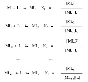
Where M and L are metal cation and ligand anion, respectively.
For the calculation of total concentration of final complex product (MLn), the overall formation constant is used.
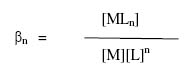
The overall formation constant is the product of stepwise formation constants
![]()
The inverse of formation constant, the dissociation constant Kd is also some time useful.

Kd had the same form as Ka for acids, which facilities comparisons between metal complexes and BrØnsted acids.
Metal complexes play an important role in various biological systems [1] and in different field of chemistry [2]. The complexation data involving essential metal ions and bioactive ligand norvaline give insight into many physicochemical processes in living systems. Banerjea has classified nickel as beneficial metal and lead as well as mercury as tonic metal in the biological systems [3]. The mercury(II), nickel(II) and lead(II) have significant biomedical applications but are toxic at higher concentration [4-27]. Norvaline an amino acid C5H11NO2 isomeric with valine and usually made synthetically. It is not found in proteins. Norvaline has several significant applications in biological systems [28-35].
Kiso [36] has done a comprehensive study on paper electrophoretic migration of metal complexes. The present electrophoretic technique is almost free from a number of defects such as temperature during electrophoresis, capillary flow on paper, electroosmosis and adsorption. The technique is very convenient in use. It gives results in fair agreement with accepted literature values. Communications [37, 38] from our laboratory described a new method for the study of metal complexes. The present work is extension of this technique and reports observations on binary systems, viz.: Hg(II) / Ni(II) / Pb(II) – norvaline.
RESULTS
The electrophoretic mobility of the metal spot against pH gives a curve with a number of plateaus as is shown in Figure 3. A constant speed over a range of pH is possible only when a particular complex species is overwhelmingly formed. Thus, every plateau is indicative of formation of a certain complex species. The first one corresponds to a region in which metal ions are uncomplexed. In this region of low pH the concentration of the [CH3 CH2 CH2 CH (NH3+) COOH] species of norvaline is at a maximum and this species is non-complexing. Beyond this range, metal ion spots have progressively decreasing mobility, complexation of metal ions should be taking place with anionic species of norvaline whose concentration increases progressively with an increase of pH. Figure 3 shows three plateaus in Hg(II), Ni(II) and Pb(II), hence all three Hg(II), Ni(II) and Pb(II) form two complexes with the norvaline anion. It is therefore assumed that the anionic species [CH3 CH2 CH2 CH(NH2) COO-] must have combined with Hg(II), Ni(II) and Pb(II) to form [Hg {CH3 CH2 CH2 CH(NH2) COO}]+, [Ni {CH3 CH2 CH2 CH(NH2) COO}]+ and [Pb {CH3 CH2 CH2 CH(NH2) COO}]+complex cations, respectively. With a further increase of pH, mobility in all three metal ions decreases giving rise to a third plateau with zero mobility that indicates its neutral nature. The third plateau in each case is due to a (1:2) metal-ligand complex. Hence, two [CH3 CH2 CH2 CH(NH2) COO-] must have combined with Hg(II), Ni(II) and Pb(II) to give the [Hg {CH3 CH2 CH2 CH(NH2) COO}2], [Ni {CH3 CH2 CH2 CH(NH2) COO}2] and [Pb{CH3 CH2 CH2 CH(NH2) COO}2] complexes, respectively.
Further increase of pH has no effect on the mobility of metal ions, which indicates no further interaction between metal ions and ligand. In general the complexation of metal ions with norvaline anion may be represented as:
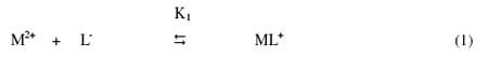
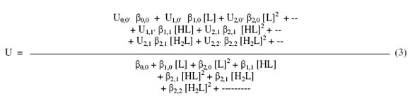
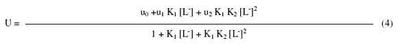
where M2+ is Hg2+ Ni2+ and Pb2+ metal ions; [L-] is the norvaline anion; K1 and K2 are the first and second stability constants, respectively. The metal spot on the paper is thus a combination of uncomplexed metal ions; 1:1 and 1:2 metal complexes. The spot is moving under the influence of electric field and the overall mobility U is given by equation (3) [39].

where u0,0 is the speed of uncomplexed metal ion, u1,0 is the speed of complex formed by the combination of one unprotonated anionic ligand with metal ion and ux·p is the speed of the metal complex formed by the combination of x anions containing, p, protons each. βs are taking the overall stability constant of the different metal complexes formed in the interaction. On taking into consideration different equilibrium above equation transformed into following useful form
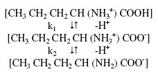
where u0, u1 and u2 are mobilities of uncomplexed metal ion, 1:1 metal complex and 1:2 metal complex, respectively. The dissociation constants of pure norvaline (k1 = 102.31; k2 = 109.65) [40] were determined by same paper electrophoretic technique. The mode of dissociation of pure norvaline can be represented as:
Using dissociation constants of pure norvaline the concentration of norvaline anion [L-] is determined for the pH value(s) of interest, from which K1 can be calculated. The concentration of complexing norvalilne anion [L-] is calculated with the help of equation
where [LT] is total concentration of ligand norvaline (0.01 M); k1 and k2 are first and second dissociation constants of pure norvaline ,respectively.

For calculating first stability constant, K1, the region between first and second plateau is pertinent. The overall mobility will be equal to the arithmetic mean of the mobility of uncomplex u0, and that of first complex, u1 at a pH value where K1 = 1/[L-].
The second stability constant, K2, of 1:2 complex can be calculated by taking into consideration, the region between second and third plateau of mobility curve. The(se) calculated value of K1 and K2 are given in Table 1.
Table 1. Stability constants of binary complexes of mercury(II), nickel(II) and lead(II) with norvaline.

DISCUSSION
It is observed from Table 1 that stability constants follow the order:
mercury(II) > nickel(II) > lead(II)
The high stability constant values of the mercury(II) – norvaline complexes indicate strong bonding between the mercury(II) cation and the norvaline anion, while the low stability constant values of lead(II) – norvaline complexes indicate weak bonding between the lead(II) cation and norvaline anion. The higher stability of mercury(II) complexes may be ascribed to the greater affinity of mercury(II) for the oxygen donor ligands. The molecular structure of norvaline given as:

It is also observed from Table 1 that the second stability constant values are found to be lower in comparison to the first stability constant in each case. This may be due to the decrease in coordinating tendency of the ligand with higher state of aggregation [41].
It is clear from the Table 1 that calculated stability constant values are approximately similar to literature values. The slight divergence in the values obtained from different sources is mainly due to the difference in temperature and ionic strength used by different sources. According to standard deviation (statistics) the precision of the method is limited to that of paper electrophoresis, and uncertainty in the result is ± 5 %. Hence, it cannot immediately replace the most reliable methods, even though it is new approach deserving further development. The proposed structure of ML2 complex is given as follows:

CONCLUDING REMARKS
Several conclusions can be drawn from the present study.
1. Mercury(II), nickel(II) and lead(II) are significant for biological systems but as such they are toxic at higher concentration.
2. Norvaline may be used to reduce the level of these metal ions in biological systems.
3. Mercury(II) – norvaline and lead(II) – norvaline complexes are found to have highest and lowest stability constant values, respectively.
4. The ML2 binary complexes are found to have lower stability constant values in comparison to ML complexes in each system.
5. The present modified electrophoretic technique is helpful in finding if complexes are formed or not, and if formed their stability constants can also be determined.
Biologically important mercury(II), nickel(II) and lead(II) complexes with norvaline can be prepared on large scale at a particular pH of the background electrolyte solution.
EXPERIMENTAL
Instruments
Systronics (Naroda, India) paper electrophoresis equipment horizontal-cum-vertical type, model 604, has been used. The apparatus consisted of a PVC moulded double tank vessel. In our laboratory significant change in the instrument has been made. Two hollow rectangular plates covered with thin polythene sheets have been used through which thermostated water is run for controlling the temperature. The tanks were closed with a transparent PVC moulded lid. The whole assembly is tight, which prevent moisture changes, which may upset the equilibria in a paper strip. This assembly design thus keeps to a minimum the disturbing effects of evaporation from the unwanted liquid flow in the paper. Each electrolyte tank contains a separate electrode chamber.
Elico (Hyderabad, India), Model L1-10, pH meter using a glass and calomel electrodes assembly working on 220 V/50 Hz established a.c. mains, was used for the pH measurements. pH meter was calibrated with buffer solution of pH 7.0. The electrophoresis cell showing sandwiched paper strips and water supply are shown in Figure 1.
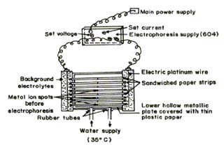
Fig.1 Electrophoresis cell showing sandwiched paper strips
Chemicals
Mercury(II), nickel(II) and lead(II) perchlorate solutions were prepared by preliminary precipitation of metal carbonates from a 0.1 M solution of sodium carbonate (AnalaR grade, BDH, Poole, UK). The precipitates were thoroughly washed with boiling water and treated with calculated amounts of 1 % perchloric acid. The resulting mixture was heated to boiling on a water bath and then filtered. The metal content of the filtrates were determined and final concentration was kept at 0.005 M [42, 43]. The position of the Ni2+ spots on the paper at the end of the experiment was detected using ammonical dimethylglyoxime (DMG), that of Pb2+ detected by 0.1% solution of 1-(2-pyridylazo) – 2- naphthol (PAN) (Merck, Darmstadt, Germany) in ethanol, that of Hg2+ detected using hydrogen sulphide in water. The 0.005 M glucose (BDH, AnalaR) solution was prepared in water and used as an indicator for the correction due to electroosmosis. A saturated aqueous solution (0.9 mL) of silver nitrate was diluted with acetone to 20 mL. Glucose was detected by spraying with this silver nitrate solution and then with 2% ethanolic solution of sodium hydroxide, when a black spot was formed. Paper strips showing the positions of the metal ion spots after electrophoresis are shown in Figure 2.
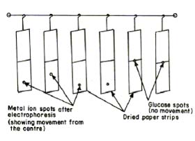
Fig.2 Paper strips showing position of metal spots after electrophoresis
Background electrolyte (BGE)
Stock solution of 5.0 M perchloric acid was prepared from its 70% solution (SDS, AnalaR grade). 2.0 M sodium hydroxide and 0.5 M norvaline (BDH, Poole, UK) solutions were prepared. The background electrolyte used in the study of binary complexes were 0.1 M perchloric acid and 0.1 M norvaline. The system was maintained at various pH by the addition of sodium hydroxide.
Procedure
Whatman No. 1 filter paper for chromatography was used for the purpose of electrophoresis. For recording observation of particular metal ion, two strips were spotted with the metal ion solution along with additional two spotted with glucose using 1.0 ml pipette and then mounted on the insulated plate. Each of the two electrolyte vessels were filled with 150 ml of background electrolyte containing 0.1 M perchloric acid and 0.01 M norvaline. The paper become moistened with the background electrolyte solutions due to diffusion. The second insulated plate was placed on paper strips and then thermostated water (35 ° C) was circulated in the plates to keep the temperature constant. The lid was then placed on the instrument to make it air tight. It was left for 10 minutes to insure wetting of strips. Subsequently a direct 200 Volts potential was applied between the electrodes. Electrophoresis was carried for 60 minutes after which these strips were removed from the tank and dried. The metal ion and glucose spots were detected by specific reagents. The leading and tailing edge were measured from the marked centre point and the mean were taken. The distance moved by glucose was subtracted (in case of migration toward anode) to obtain correct path length. Migration towards anode and cathode were designated by negative and positive signs ,respectively.
Electrophoretic observations on metal ions were recorded at various pH values of the background electrolyte obtained by adding NaOH solution. The ionic strength being maintained at 0.1 M. The observed mobility of migrant was calculated by using the formula.
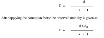
where U = mobility of metal ion / complex ion; d = mean of duplicate distance travelled by metal ion / complex ion; dG = mean of duplicate distance travelled by glucose spot; x = field strength; t = time for electrophoresis.
The protonation constants of pure norvaline were determined by same paper electrophoresis technique. The two paper strips were spotted with pure norvaline along with two with glucose using 0.1 M perchloric acid only in a background electrolyte. The electrophoresis was carried for 60 minutes as for metal ions. The electrophoretic speed was calculated. The speed of the metal ion / norvaline spots are reported with pH values. The individual speeds of the duplicate spots were found to be fairly equal.
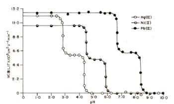
Fig. 3 Mobility curves for the metal(II) – norvaline systems. ![]() = Hg(II) –norvaline; = nickel(II) – norvaline
= Hg(II) –norvaline; = nickel(II) – norvaline ![]()
![]() = Pb(II) – norvaline. Background electrolyte = 0.1 M perchloric acid and 0.01 M norvaline. pH was maintained by addition of sodium hydroxide. Concentration of Hg2+, Ni2+ and Pb2+= 0.005 M. Ionic strength = 0.1 M. Temperature = 35 °C. The paper strips were spotted with 0.1 ml of sample solutions and glucose (for making osmotic corrections).
= Pb(II) – norvaline. Background electrolyte = 0.1 M perchloric acid and 0.01 M norvaline. pH was maintained by addition of sodium hydroxide. Concentration of Hg2+, Ni2+ and Pb2+= 0.005 M. Ionic strength = 0.1 M. Temperature = 35 °C. The paper strips were spotted with 0.1 ml of sample solutions and glucose (for making osmotic corrections).
REFERENCES
1. SIGEL H., Metal Ions in Biological Systems, Marcel Dekker, New York, 1989, Vols. 1-23. [ Links ]
2. SHERMAN S. E., LIPPARD S. J., Chem. Rev., 1987, 87, 1153. [ Links ]
3. BENERJEA D., Everymans Sci., 1995, 29, 176. [ Links ]
4.BHARARA M. S., PERKIN S., ATWOOD D. A., Inorg. Chem., 2006, 45(18), 7261. [ Links ]
5.XIE L., FLIPPIN, J. L., DEIGHTON N., FUNK D. H., DICKEY D. A., BUCHWALTER, D. B., Environ. Sci. Technol., 2009, 43, 934. [ Links ]
6. SUTTON D. J., TCHOUNWOU P. W., Int. J. Environ. Res. Public Health, 2007, 4(2), 138. [ Links ]
7.KARTHIKEYAN J., PARAMESHWARA P., SHETTY A. N., Indian J. Chem. Technol., 2008, 13, 493. [ Links ]
8.HALBACH S., VOGT S., KOHLER W., FELGENHAUER N., WELZL G., KREMERS L., ZILKER T., MERCHART D., Environ. Res., 2008, 107, 69. [ Links ]
9.WAGNER – DÖBLER L., Appl. Microbiol. Biotechnol. ,2003, 62, 124. [ Links ]
10.SUMI D., J. Health Sci., 2008, 54(3), 267. [ Links ]
11.KUMAR G., RAI P., Turk J. Biol., 2007, 31, 13. [ Links ]
12. SAEED S., RASHID N., ALI M., HUSSAIN R., Eur. J. Chem., 2010, 1(3), 2000. [ Links ]
13. ALY A. A. M., OSMAN A. H., ELMOTTALEB M., GOUDA G. A. H., J. Chil. Chem. Soc., 2009, 54(4), 349. [ Links ]
14.CHANDRA S., TYAGI M., SHARMA K., J. Iran. Chem. Soc., 2009, 6(2), 310. [ Links ]
15. KHAN M. R., KHAN M. M., Aust. J. Basic Appl. Sci., 2010, 4(6), 1036. [ Links ]
16. MANSOOR M. M., Int. J. Chem Tech. Res., 2010, 2(1), 640. [ Links ]
17. NAALIYA J., Bayero J. Pure Appl. Sci., 2010, 3(1), 241. [ Links ]
18. BULUT I., Turk. J. Chem. 2009, 33, 507. [ Links ]
19. CHIPASA K. B., Waste Management, 2003, 23, 135. [ Links ]
20. ADEYEMI A. O., Int. J. Environ. Res., 2009, 3(4), 477. [ Links ]
21. LEVINA V. V., NOLEN V., SU Y., GODWIN A. K., FISHMAN D., HERBERMAN R. B., LOKSHIN A. E., Cancer Res., 2009, 69(12), 5226. [ Links ]
22. VANYSEK P., RAMIREZ L. B., J. Chil. Chem. Soc., 2008, 53(2), 1455. [ Links ]
23. WU C., XIAOYONG Z., TIAN Y., SONG J., YANG D., ROGGENDORF M., CHEN X., LU M., J. Gen. Virol. ,2010, 91, 483. [ Links ]
24. DAVID S. S., MEGGERS E., Curr. Opin. Chem. Biol., 2008, 12(2), 194. [ Links ]
25. IMANPOOR M. R., BAGHERI T., World J. Zoology, 2010, 5(4), 278. [ Links ]
26. ADEBAYO O. L., ADEGBESAN B. O., ADENUGO G.A ., Asian J. Biol. Sci., 2009, 2(1), 7. [ Links ]
27. SILVEIRA E. A., LIZARDO J. H. F., SOUZA L. P., STEFANAN I., VASSALLO, Braz. J. Med. Biol. Res., 2010, 43(6), 492. [ Links ]
28. ZANI M.-L., BARANGER K., GUYOT N., DALLET-CHOISY S., MOREAU T., Protein Sci., 2009, 18(3), 579. [ Links ]
29. SIT C. S., VEDERAS J. C., Biochem. Cell Biol., 2008, 86(2), 116. [ Links ]
30. CAUPENE C., CHAUME G., RICARD L., BRIGAUD T., Org. Lett., 2009, 11(1), 209. [ Links ]
31. CHEN K. X., NJOROGE G., RICHARDO J., PRONGAY A., BUTKIEWICZ, N., YAO N., MADISON V., GIRIJAVALLABHAN V., J. Med. Chem. 2005, 48(20), 6229. [ Links ]
32. LEE J., FINLEY J. W., HARNLY J. M., J. Agri. Food Chem., 2005, 53(23), 9105. [ Links ]
33. BERGSENG E., XIA, J., KHOSLA C., SOLLID L. M., J. Biol. Chem., 2005, 280, 21791. [ Links ]
34. BIALAS A., KAFARSKI P., Curr. Med. Chem., 2009, 9(7), 728. [ Links ]
35. SANKARANARAYANAN R., CHERNEY M. M., CHERNEY L. T., GAREN C. R., MORADIAN F., MICHAL N. C., J. Mol. Biol., 2008, 25(4), 1052. [ Links ]
36. KISO Y., Electrophoresis - New Attempts of Ionics, Nankodo, Japan, 1972. [ Links ]
37. TEWARI B. B., Russ J. Coord. Chem., 2003, 29, 441. [ Links ]
38. TEWARI B. B., J. Mex. Chem. Soc., 2008, 52(3), 222. [ Links ]
39. JOKL, V., J. Chromatogr.,1964, 6, 432. [ Links ]
40. MARTELL A. E., SMITH R. M., Critical Stability Constants, Amino Acids, Plenum Press, New York, 1974, 1, 7. [ Links ]
41. JOSHI J. D., BHATTACHARYA P. K., J. Indian Chem. Soc., 1980, 57, 336. [ Links ]
42. KOLTHOFF I. M., BELCHER R., Volumetric Analysis, Vol. 3, Interscience Publishers Inc., New York, USA, 1957. [ Links ]
43. Vogel A. I., Text Book of Quantitative Inorganic Analysis, including Elementary Instrumental Analysis, 4th Edition, Longmans, London, UK, 1978. [ Links ]
44. PERRIN D. D., Stability Constants of Metal Ion Complexes, Part B, Organic Ligands, Pergamon Press, Oxford, UK, IUPAC Series No. 22, 1979, P. 325. [ Links ]
45. SILLEN L. G., MARTELL A. E., Stability Constants of Metal Ion Complexes, Special Publication No. 17, Chemical Society London, 1974, pp. 462-463. [ Links ]

